Movie
Movie Controller

[English] 日本語
 Yorodumi
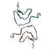
- PDB-9c5r: Structure of recombinantly assembled alpha-synuclein variant fibrils -
+ Open data

- Basic information
| Entry | Database: PDB / ID: 9c5r | |||||||||
|---|---|---|---|---|---|---|---|---|---|---|
| Title | Structure of recombinantly assembled alpha-synuclein variant fibrils | |||||||||
 Components Components | Alpha-synuclein | |||||||||
 Keywords Keywords | PROTEIN FIBRIL / alpha-synuclein fibrils / structural protein | |||||||||
| Function / homology |  Function and homology information Function and homology informationnegative regulation of mitochondrial electron transport, NADH to ubiquinone / neutral lipid metabolic process / regulation of acyl-CoA biosynthetic process / negative regulation of dopamine uptake involved in synaptic transmission / negative regulation of norepinephrine uptake / response to desipramine / positive regulation of SNARE complex assembly / positive regulation of hydrogen peroxide catabolic process / supramolecular fiber / regulation of synaptic vesicle recycling ...negative regulation of mitochondrial electron transport, NADH to ubiquinone / neutral lipid metabolic process / regulation of acyl-CoA biosynthetic process / negative regulation of dopamine uptake involved in synaptic transmission / negative regulation of norepinephrine uptake / response to desipramine / positive regulation of SNARE complex assembly / positive regulation of hydrogen peroxide catabolic process / supramolecular fiber / regulation of synaptic vesicle recycling / negative regulation of chaperone-mediated autophagy / regulation of reactive oxygen species biosynthetic process / positive regulation of protein localization to cell periphery / mitochondrial membrane organization / negative regulation of exocytosis / regulation of glutamate secretion / dopamine biosynthetic process / positive regulation of neurotransmitter secretion / response to iron(II) ion / regulation of macrophage activation / negative regulation of dopamine metabolic process / negative regulation of platelet-derived growth factor receptor signaling pathway / SNARE complex assembly / negative regulation of thrombin-activated receptor signaling pathway / Lewy body / regulation of locomotion / negative regulation of microtubule polymerization / synaptic vesicle priming / regulation of norepinephrine uptake / transporter regulator activity / protein kinase inhibitor activity / positive regulation of inositol phosphate biosynthetic process / synaptic vesicle transport / dopamine uptake involved in synaptic transmission / regulation of dopamine secretion / positive regulation of receptor recycling / cuprous ion binding / positive regulation of exocytosis / nuclear outer membrane / mitochondrial ATP synthesis coupled electron transport / dynein complex binding / synaptic vesicle exocytosis / response to magnesium ion / positive regulation of endocytosis / negative regulation of serotonin uptake / response to type II interferon / cysteine-type endopeptidase inhibitor activity / kinesin binding / regulation of presynapse assembly / synaptic vesicle endocytosis / alpha-tubulin binding / beta-tubulin binding / phospholipase binding / phospholipid metabolic process / supramolecular fiber organization / behavioral response to cocaine / cellular response to fibroblast growth factor stimulus / cellular response to epinephrine stimulus / inclusion body / Hsp70 protein binding / enzyme inhibitor activity / response to interleukin-1 / axon terminus / cellular response to copper ion / regulation of microtubule cytoskeleton organization / positive regulation of release of sequestered calcium ion into cytosol / SNARE binding / adult locomotory behavior / glutathione metabolic process / protein tetramerization / protein sequestering activity / tubulin binding / phosphoprotein binding / excitatory postsynaptic potential / microglial cell activation / ferrous iron binding / fatty acid metabolic process / phospholipid binding / PKR-mediated signaling / synapse organization / receptor internalization / regulation of long-term neuronal synaptic plasticity / protein destabilization / tau protein binding / enzyme activator activity / positive regulation of inflammatory response / terminal bouton / long-term synaptic potentiation / synaptic vesicle membrane / actin cytoskeleton / growth cone / actin binding / cellular response to oxidative stress / neuron apoptotic process / cell cortex / histone binding / response to lipopolysaccharide / microtubule binding / amyloid fibril formation / chemical synaptic transmission Similarity search - Function | |||||||||
| Biological species |  Homo sapiens (human) Homo sapiens (human) | |||||||||
| Method | ELECTRON MICROSCOPY / helical reconstruction / cryo EM / Resolution: 2.61 Å | |||||||||
 Authors Authors | Sun, C.Q. / Zhou, K. / DePaola IV, P. / Lee, V.M.Y. / Li, C. / Zhou, Z.H. / Rodriguez, J.A. / Peng, C. / Jiang, L. | |||||||||
| Funding support |  United States, 2items United States, 2items
| |||||||||
 Citation Citation | Journal: J Biol Chem / Year: 2025 Title: Structural basis of a distinct α-synuclein strain that promotes tau inclusion in neurons. Authors: Chuanqi Sun / Kang Zhou / Peter DePaola / Cally Li / Virginia M Y Lee / Z Hong Zhou / Chao Peng / Lin Jiang / Abstract: Amyloidoses are predominantly associated with the accumulation of persistent aggregates of a particular protein. For example, the protein α-synuclein characteristically aggregates in Parkinson's ...Amyloidoses are predominantly associated with the accumulation of persistent aggregates of a particular protein. For example, the protein α-synuclein characteristically aggregates in Parkinson's disease (PD), while amyloid beta and tau deposits are associated with Alzheimer's disease (AD). However, α-synuclein-positive inclusions have been reportedly found in some tauopathies, and vice versa; tau-positive inclusions can be found in synucleinopathies. This suggests that there may be coexistence or crosstalk between these proteinopathies. This coexistence suggests that the simultaneous presence of these misfolded proteins may amplify pathogenic mechanisms. However, the crosstalk between these two types of proteopathies remains poorly understood. We now determine the structure of α-synuclein fibrils that directly promote tau aggregation by cryogenic electron microscopy. Helical reconstruction at 2.6 Å resolution reveals a new α-synuclein fibril polymorph we term "strain B"; its core is unique, incorporating both the N- and C-termini of α-synuclein. The design of peptides meant to inhibit the formation of this structure demonstrates that the C-terminal domain fragment (D105-E115) of α-synuclein is critical for the formation of "strain B" fibrils and may play a key role in its interaction with tau. We hypothesize that the unique structure of pathological α-synuclein significantly contributes to tau co-aggregation and plays a role in the intricate interactions among Alzheimer's, Parkinson's, and other neurodegenerative diseases. These findings open new avenues for drug targeting, discovery, and improve our understanding of neurodegenerative pathology. | |||||||||
| History |
|
- Structure visualization
| Structure viewer | Molecule: MolmilJmol/JSmol |
|---|
- Downloads & links
-Download
| PDBx/mmCIF format | 9c5r.cif.gz | 138.6 KB | Display | PDBx/mmCIF format |
|---|---|---|---|---|
| PDB format | pdb9c5r.ent.gz | 108.9 KB | Display | PDB format |
| PDBx/mmJSON format | 9c5r.json.gz | Tree view | PDBx/mmJSON format | |
| Others |  Other downloads Other downloads |
-Validation report
| Arichive directory | https://data.pdbj.org/pub/pdb/validation_reports/c5/9c5rftp://data.pdbj.org/pub/pdb/validation_reports/c5/9c5r | HTTPS FTP |
|---|
-Related structure data
| Related structure data |  45221MC M: map data used to model this data C: citing same article ( |
|---|---|
| Similar structure data |
-Links
 PDBj
PDBj- Assembly
| Deposited unit | 
|
|---|---|
| 1 |
|
| Symmetry | Helical symmetry: (Circular symmetry: 1 / Dyad axis: no / N subunits divisor: 1 / Num. of operations: 10 / Rise per n subunits: 2.408 Å / Rotation per n subunits: 179.477 °) |
-Components
| #1: Protein | Mass: 14476.108 Da / Num. of mol.: 10 Source method: isolated from a genetically manipulated source Source: (gene. exp.) Homo sapiens (human) / Gene: SNCA, NACP, PARK1Production host:  References: UniProt: P37840 Has protein modification | N | |
|---|
-Experimental details
-Experiment
| Experiment | Method: ELECTRON MICROSCOPY |
|---|---|
| EM experiment | Aggregation state: FILAMENT / 3D reconstruction method: helical reconstruction |
- Sample preparation
| Component | Name: alpha-synuclein / Type: COMPLEX / Entity ID: all / Source: RECOMBINANT | ||||||||||||||||||||
|---|---|---|---|---|---|---|---|---|---|---|---|---|---|---|---|---|---|---|---|---|---|
| Molecular weight | Value: 60 kDa/nm / Experimental value: NO | ||||||||||||||||||||
| Source (natural) | Organism: Homo sapiens (human) | ||||||||||||||||||||
| Source (recombinant) | Organism: | ||||||||||||||||||||
| Buffer solution | pH: 7.5 | ||||||||||||||||||||
| Buffer component |
| ||||||||||||||||||||
| Specimen | Conc.: 8 mg/ml / Embedding applied: NO / Shadowing applied: NO / Staining applied: NO / Vitrification applied: YES | ||||||||||||||||||||
| Specimen support | Grid material: COPPER / Grid mesh size: 300 divisions/in. / Grid type: Quantifoil R2/1 | ||||||||||||||||||||
| Vitrification | Instrument: FEI VITROBOT MARK IV / Cryogen name: ETHANE / Humidity: 100 % / Chamber temperature: 283.15 K |
- Electron microscopy imaging
| Experimental equipment |  Model: Titan Krios / Image courtesy: FEI Company |
|---|---|
| Microscopy | Model: FEI TITAN KRIOS |
| Electron gun | Electron source:  FIELD EMISSION GUN / Accelerating voltage: 300 kV / Illumination mode: FLOOD BEAM FIELD EMISSION GUN / Accelerating voltage: 300 kV / Illumination mode: FLOOD BEAM |
| Electron lens | Mode: BRIGHT FIELD / Nominal magnification: 81000 X / Nominal defocus max: 2200 nm / Nominal defocus min: 900 nm / Calibrated defocus min: 700 nm / Calibrated defocus max: 2800 nm / Cs: 2.7 mm / C2 aperture diameter: 50 µm / Alignment procedure: COMA FREE |
| Specimen holder | Cryogen: NITROGEN / Specimen holder model: FEI TITAN KRIOS AUTOGRID HOLDER |
| Image recording | Average exposure time: 3.6 sec. / Electron dose: 48 e/Å2 / Detector mode: SUPER-RESOLUTION / Film or detector model: GATAN K3 (6k x 4k) / Num. of grids imaged: 1 / Num. of real images: 4094 |
| EM imaging optics | Energyfilter name: GIF Quantum LS / Energyfilter slit width: 20 eV |
| Image scans | Width: 5760 / Height: 4092 / Movie frames/image: 5 / Used frames/image: 2-34 |
- Processing
| EM software |
| ||||||||||||||||||||||||||||||||||||
|---|---|---|---|---|---|---|---|---|---|---|---|---|---|---|---|---|---|---|---|---|---|---|---|---|---|---|---|---|---|---|---|---|---|---|---|---|---|
| CTF correction | Type: PHASE FLIPPING AND AMPLITUDE CORRECTION | ||||||||||||||||||||||||||||||||||||
| Helical symmerty | Angular rotation/subunit: 179.477 ° / Axial rise/subunit: 2.408 Å / Axial symmetry: C1 | ||||||||||||||||||||||||||||||||||||
| Particle selection | Num. of particles selected: 57074 | ||||||||||||||||||||||||||||||||||||
| 3D reconstruction | Resolution: 2.61 Å / Resolution method: FSC 0.143 CUT-OFF / Num. of particles: 26051 / Algorithm: FOURIER SPACE / Num. of class averages: 1 / Symmetry type: HELICAL | ||||||||||||||||||||||||||||||||||||
| Atomic model building | B value: 53.18 / Protocol: RIGID BODY FIT / Space: REAL | ||||||||||||||||||||||||||||||||||||
| Atomic model building | PDB-ID: 6CU7 Pdb chain-ID: A / Accession code: 6CU7 / Source name: PDB / Type: experimental model |