National Institutes of Health/National Institute on Aging (NIH/NIA)
R01 AG060149
United States
National Institutes of Health/National Institute of General Medical Sciences (NIH/NIGMS)
R01 GM071940
United States
Citation
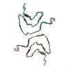
Journal: J Biol Chem / Year: 2025 Title: Structural basis of a distinct α-synuclein strain that promotes tau inclusion in neurons. Authors: Chuanqi Sun / Kang Zhou / Peter DePaola / Cally Li / Virginia M Y Lee / Z Hong Zhou / Chao Peng / Lin Jiang / Abstract: Amyloidoses are predominantly associated with the accumulation of persistent aggregates of a particular protein. For example, the protein α-synuclein characteristically aggregates in Parkinson's ...Amyloidoses are predominantly associated with the accumulation of persistent aggregates of a particular protein. For example, the protein α-synuclein characteristically aggregates in Parkinson's disease (PD), while amyloid beta and tau deposits are associated with Alzheimer's disease (AD). However, α-synuclein-positive inclusions have been reportedly found in some tauopathies, and vice versa; tau-positive inclusions can be found in synucleinopathies. This suggests that there may be coexistence or crosstalk between these proteinopathies. This coexistence suggests that the simultaneous presence of these misfolded proteins may amplify pathogenic mechanisms. However, the crosstalk between these two types of proteopathies remains poorly understood. We now determine the structure of α-synuclein fibrils that directly promote tau aggregation by cryogenic electron microscopy. Helical reconstruction at 2.6 Å resolution reveals a new α-synuclein fibril polymorph we term "strain B"; its core is unique, incorporating both the N- and C-termini of α-synuclein. The design of peptides meant to inhibit the formation of this structure demonstrates that the C-terminal domain fragment (D105-E115) of α-synuclein is critical for the formation of "strain B" fibrils and may play a key role in its interaction with tau. We hypothesize that the unique structure of pathological α-synuclein significantly contributes to tau co-aggregation and plays a role in the intricate interactions among Alzheimer's, Parkinson's, and other neurodegenerative diseases. These findings open new avenues for drug targeting, discovery, and improve our understanding of neurodegenerative pathology.
In the structure databanks used in Yorodumi, some data are registered as the other names, "COVID-19 virus" and "2019-nCoV". Here are the details of the virus and the list of structure data.
Jan 31, 2019. EMDB accession codes are about to change! (news from PDBe EMDB page)
EMDB accession codes are about to change! (news from PDBe EMDB page)
The allocation of 4 digits for EMDB accession codes will soon come to an end. Whilst these codes will remain in use, new EMDB accession codes will include an additional digit and will expand incrementally as the available range of codes is exhausted. The current 4-digit format prefixed with “EMD-” (i.e. EMD-XXXX) will advance to a 5-digit format (i.e. EMD-XXXXX), and so on. It is currently estimated that the 4-digit codes will be depleted around Spring 2019, at which point the 5-digit format will come into force.
The EM Navigator/Yorodumi systems omit the EMD- prefix.
Related info.:Q: What is EMD? / ID/Accession-code notation in Yorodumi/EM Navigator
Yorodumi is a browser for structure data from EMDB, PDB, SASBDB, etc.
This page is also the successor to EM Navigator detail page, and also detail information page/front-end page for Omokage search.
The word "yorodu" (or yorozu) is an old Japanese word meaning "ten thousand". "mi" (miru) is to see.
Related info.:EMDB / PDB / SASBDB / Comparison of 3 databanks / Yorodumi Search / Aug 31, 2016. New EM Navigator & Yorodumi / Yorodumi Papers / Jmol/JSmol / Function and homology information / Changes in new EM Navigator and Yorodumi
 Movie
Movie Controller

 Yorodumi
 Open data
 Basic information
 Map data
 Sample
 Keywords
 Function and homology information
 Homo sapiens (human)
 Authors
 United States, 2 items
  Citation
 Structure visualization
 Downloads & links
 emd_45221.png
 http://ftp.pdbj.org/pub/emdb/structures/EMD-45221
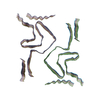
 Z (Sec.)
 Y (Row.)
 X (Col.)


 Sample components
 Processing
 Electron microscopy
 FIELD EMISSION GUN