 Movie
Movie Controller

[English] 日本語
 Yorodumi
- PDB-9bsx: Cryo-EM structure of HCoV-HKU1 glycoprotein in complex with 9O-ac... -
+ Open data

- Basic information
| Entry | Database: PDB / ID: 9bsx | ||||||
|---|---|---|---|---|---|---|---|
| Title | Cryo-EM structure of HCoV-HKU1 glycoprotein in complex with 9O-acetyl GD3 sialoglycan (DAA state) | ||||||
 Components Components | Spike glycoprotein | ||||||
 Keywords Keywords | VIRAL PROTEIN / HCoV-HKU1 spike glycoprotein ectodomain / proline stablized | ||||||
| Function / homology |  Function and homology information Function and homology informationhost cell endoplasmic reticulum-Golgi intermediate compartment membrane / receptor-mediated virion attachment to host cell / endocytosis involved in viral entry into host cell / fusion of virus membrane with host plasma membrane / fusion of virus membrane with host endosome membrane / viral envelope / host cell plasma membrane / virion membrane / membrane Similarity search - Function | ||||||
| Biological species |  Human coronavirus HKU1 Human coronavirus HKU1 | ||||||
| Method | ELECTRON MICROSCOPY / single particle reconstruction / cryo EM / Resolution: 2.37 Å | ||||||
 Authors Authors | Jin, M. / Rini, J.M. | ||||||
| Funding support |  Canada, 1items Canada, 1items
| ||||||
 Citation Citation | Journal: Nat Commun / Year: 2025 Title: Human coronavirus HKU1 spike structures reveal the basis for sialoglycan specificity and carbohydrate-promoted conformational changes. Authors: Min Jin / Zaky Hassan / Zhijie Li / Ying Liu / Aleksandra Marakhovskaia / Alan H M Wong / Adam Forman / Mark Nitz / Michel Gilbert / Hai Yu / Xi Chen / James M Rini /  Abstract: The human coronavirus HKU1 uses both sialoglycoconjugates and the protein transmembrane serine protease 2 (TMPRSS2) as receptors. Carbohydrate binding leads to the spike protein up conformation ...The human coronavirus HKU1 uses both sialoglycoconjugates and the protein transmembrane serine protease 2 (TMPRSS2) as receptors. Carbohydrate binding leads to the spike protein up conformation required for TMPRSS2 binding, an outcome suggesting a distinct mechanism for driving fusion of the viral and host cell membranes. Nevertheless, the conformational changes promoted by carbohydrate binding have not been fully elucidated and the basis for HKU1's carbohydrate binding specificity remains unknown. Reported here are high resolution cryo-EM structures of the HKU1 spike protein trimer in its apo form and in complex with the carbohydrate moiety of a candidate carbohydrate receptor, the 9-O-acetylated GD3 ganglioside. The structures show that the spike monomer can exist in four discrete conformational states and that progression through them would promote the up conformation upon carbohydrate binding. We also show that a six-amino-acid insert is a determinant of HKU1's specificity for gangliosides containing a 9-O-acetylated α2-8-linked disialic acid moiety and that HKU1 shows weak affinity for the 9-O-acetylated sialic acids found on decoy receptors such as mucins. | ||||||
| History |
|
- Structure visualization
| Structure viewer | Molecule: MolmilJmol/JSmol |
|---|
- Downloads & links
-Download
| PDBx/mmCIF format | 9bsx.cif.gz | 754.6 KB | Display | PDBx/mmCIF format |
|---|---|---|---|---|
| PDB format | pdb9bsx.ent.gz | Display | PDB format | |
| PDBx/mmJSON format | 9bsx.json.gz | Tree view | PDBx/mmJSON format | |
| Others |  Other downloads Other downloads |
-Validation report
| Arichive directory | https://data.pdbj.org/pub/pdb/validation_reports/bs/9bsxftp://data.pdbj.org/pub/pdb/validation_reports/bs/9bsx | HTTPS FTP |
|---|
-Related structure data
| Related structure data |  44875MC  9bswC  9bsyC  9bszC  9bt0C  9bt1C  9bt2C  9bt9C  9btaC 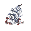 9btbC  9btcC  9btdC  9n12C  9n13C  9n14C  9n15C  9n16C  9n17C  9n18C  9n19C M: map data used to model this data C: citing same article ( |
|---|---|
| Similar structure data |


-Links
 PDBj
PDBj- Assembly
| Deposited unit | 
|
|---|---|
| 1 |
|
-Components
-Protein / Non-polymers , 2 types, 6 molecules ABC
| #1: Protein | Mass: 150632.734 Da / Num. of mol.: 3 / Fragment: ectodomain (UNP residues 14-1298) Source method: isolated from a genetically manipulated source Source: (gene. exp.) Human coronavirus HKU1 / Gene: S, 3 / Production host:  Homo sapiens (human) / References: UniProt: Q5MQD0 Homo sapiens (human) / References: UniProt: Q5MQD0#6: Chemical | Mass: 642.561 Da / Num. of mol.: 3 / Source method: obtained synthetically / Formula: C24H38N2O18 / Feature type: SUBJECT OF INVESTIGATION |
|---|
-Sugars , 4 types, 62 molecules 
| #2: Polysaccharide | Source method: isolated from a genetically manipulated source #3: Polysaccharide | 2-acetamido-2-deoxy-beta-D-glucopyranose-(1-4)-2-acetamido-2-deoxy-beta-D-glucopyranose Source method: isolated from a genetically manipulated source #4: Polysaccharide | Source method: isolated from a genetically manipulated source #5: Sugar | ChemComp-NAG /  Type: D-saccharide, beta linking / Mass: 221.208 Da / Num. of mol.: 39 / Source method: obtained synthetically / Formula: C8H15NO6 Type: D-saccharide, beta linking / Mass: 221.208 Da / Num. of mol.: 39 / Source method: obtained synthetically / Formula: C8H15NO6 |
|---|
-Details
| Has ligand of interest | Y |
|---|---|
| Has protein modification | Y |
-Experimental details
-Experiment
| Experiment | Method: ELECTRON MICROSCOPY |
|---|---|
| EM experiment | Aggregation state: PARTICLE / 3D reconstruction method: single particle reconstruction |
- Sample preparation
| Component | Name: Human coronavirus HKU1 spike glycoprotein / Type: COMPLEX Details: Ectodomain generated by recombinant expression in HEK293 Freestyle cells Entity ID: #1 / Source: RECOMBINANT | |||||||||||||||
|---|---|---|---|---|---|---|---|---|---|---|---|---|---|---|---|---|
| Molecular weight | Value: 0.457593 MDa / Experimental value: NO | |||||||||||||||
| Source (natural) | Organism: Human coronavirus HKU1 | |||||||||||||||
| Source (recombinant) | Organism: Homo sapiens (human) | |||||||||||||||
| Details of virus | Type: VIRION | |||||||||||||||
| Buffer solution | pH: 8 | |||||||||||||||
| Buffer component |
| |||||||||||||||
| Specimen | Conc.: 1.15 mg/ml / Embedding applied: NO / Shadowing applied: NO / Staining applied: NO / Vitrification applied: YES | |||||||||||||||
| Specimen support | Grid type: C-flat-2/2 | |||||||||||||||
| Vitrification | Instrument: FEI VITROBOT MARK IV / Cryogen name: ETHANE / Humidity: 100 % / Chamber temperature: 277 K |
- Electron microscopy imaging
| Experimental equipment |  Model: Titan Krios / Image courtesy: FEI Company |
|---|---|
| Microscopy | Model: FEI TITAN KRIOS |
| Electron gun | Electron source:  FIELD EMISSION GUN / Accelerating voltage: 300 kV / Illumination mode: FLOOD BEAM FIELD EMISSION GUN / Accelerating voltage: 300 kV / Illumination mode: FLOOD BEAM |
| Electron lens | Mode: BRIGHT FIELD / Nominal magnification: 75000 X / Nominal defocus max: 1800 nm / Nominal defocus min: 1200 nm / Cs: 2.7 mm |
| Specimen holder | Cryogen: NITROGEN / Specimen holder model: FEI TITAN KRIOS AUTOGRID HOLDER |
| Image recording | Electron dose: 36 e/Å2 / Film or detector model: FEI FALCON IV (4k x 4k) |
- Processing
| EM software |
| |||||||||
|---|---|---|---|---|---|---|---|---|---|---|
| CTF correction | Type: PHASE FLIPPING AND AMPLITUDE CORRECTION | |||||||||
| Symmetry | Point symmetry: C1 (asymmetric) | |||||||||
| 3D reconstruction | Resolution: 2.37 Å / Resolution method: FSC 0.143 CUT-OFF / Num. of particles: 307395 / Symmetry type: POINT | |||||||||
| Atomic model building | Protocol: FLEXIBLE FIT |
