 Movie
Movie Controller

[English] 日本語
 Yorodumi
- EMDB-48807: Cryo-EM structure of HCoV-HKU1 glycoprotein (Deletion 33Pro34Arg) -
+ Open data

- Basic information
| Entry |  | |||||||||
|---|---|---|---|---|---|---|---|---|---|---|
| Title | Cryo-EM structure of HCoV-HKU1 glycoprotein (Deletion 33Pro34Arg) | |||||||||
 Map data Map data | ||||||||||
 Sample Sample |
| |||||||||
 Keywords Keywords | HCoV-HKU1 spike glycoprotein ectodomain / proline stablized / VIRAL PROTEIN | |||||||||
| Function / homology |  Function and homology information Function and homology informationhost cell endoplasmic reticulum-Golgi intermediate compartment membrane / receptor-mediated virion attachment to host cell / endocytosis involved in viral entry into host cell / fusion of virus membrane with host plasma membrane / fusion of virus membrane with host endosome membrane / viral envelope / host cell plasma membrane / virion membrane / membrane Similarity search - Function | |||||||||
| Biological species |  Human coronavirus HKU1 Human coronavirus HKU1 | |||||||||
| Method | single particle reconstruction / cryo EM / Resolution: 3.01 Å | |||||||||
 Authors Authors | Jin M / Rini JM | |||||||||
| Funding support |  Canada, 1 items Canada, 1 items
| |||||||||
 Citation Citation | Journal: Nat Commun / Year: 2025 Title: Human coronavirus HKU1 spike structures reveal the basis for sialoglycan specificity and carbohydrate-promoted conformational changes. Authors: Min Jin / Zaky Hassan / Zhijie Li / Ying Liu / Aleksandra Marakhovskaia / Alan H M Wong / Adam Forman / Mark Nitz / Michel Gilbert / Hai Yu / Xi Chen / James M Rini /  Abstract: The human coronavirus HKU1 uses both sialoglycoconjugates and the protein transmembrane serine protease 2 (TMPRSS2) as receptors. Carbohydrate binding leads to the spike protein up conformation ...The human coronavirus HKU1 uses both sialoglycoconjugates and the protein transmembrane serine protease 2 (TMPRSS2) as receptors. Carbohydrate binding leads to the spike protein up conformation required for TMPRSS2 binding, an outcome suggesting a distinct mechanism for driving fusion of the viral and host cell membranes. Nevertheless, the conformational changes promoted by carbohydrate binding have not been fully elucidated and the basis for HKU1's carbohydrate binding specificity remains unknown. Reported here are high resolution cryo-EM structures of the HKU1 spike protein trimer in its apo form and in complex with the carbohydrate moiety of a candidate carbohydrate receptor, the 9-O-acetylated GD3 ganglioside. The structures show that the spike monomer can exist in four discrete conformational states and that progression through them would promote the up conformation upon carbohydrate binding. We also show that a six-amino-acid insert is a determinant of HKU1's specificity for gangliosides containing a 9-O-acetylated α2-8-linked disialic acid moiety and that HKU1 shows weak affinity for the 9-O-acetylated sialic acids found on decoy receptors such as mucins. | |||||||||
| History |
|
- Structure visualization
| Supplemental images |
|---|

- Downloads & links
-EMDB archive
| Map data | emd_48807.map.gz | 32.3 MB | EMDB map data format | |
|---|---|---|---|---|
| Header (meta data) | emd-48807-v30.xmlemd-48807.xml | 18.2 KB 18.2 KB | Display Display | EMDB header |
| FSC (resolution estimation) | emd_48807_fsc.xml | 8.4 KB | Display | FSC data file |
| Images |  emd_48807.png emd_48807.png | 50.3 KB | ||
| Masks | emd_48807_msk_1.map | 64 MB | Mask map | |
| Filedesc metadata | emd-48807.cif.gz | 7.1 KB | ||
| Others | emd_48807_half_map_1.map.gzemd_48807_half_map_2.map.gz | 59.3 MB 59.3 MB | ||
| Archive directory |  http://ftp.pdbj.org/pub/emdb/structures/EMD-48807ftp://ftp.pdbj.org/pub/emdb/structures/EMD-48807 http://ftp.pdbj.org/pub/emdb/structures/EMD-48807ftp://ftp.pdbj.org/pub/emdb/structures/EMD-48807 | HTTPS FTP |
-Related structure data
| Related structure data |  9n16MC  9bswC  9bsxC  9bsyC  9bszC  9bt0C  9bt1C  9bt2C  9bt9C  9btaC 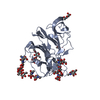 9btbC  9btcC  9btdC  9n12C  9n13C  9n14C  9n15C  9n17C  9n18C  9n19C M: atomic model generated by this map C: citing same article ( |
|---|---|
| Similar structure data |


-Links
| EMDB pages | EMDB (EBI/PDBe) / EMDataResource |
|---|
-Map
| File | Download / File: emd_48807.map.gz / Format: CCP4 / Size: 64 MB / Type: IMAGE STORED AS FLOATING POINT NUMBER (4 BYTES) | ||||||||||||||||||||||||||||||||||||
|---|---|---|---|---|---|---|---|---|---|---|---|---|---|---|---|---|---|---|---|---|---|---|---|---|---|---|---|---|---|---|---|---|---|---|---|---|---|
| Projections & slices | Image control
Images are generated by Spider. | ||||||||||||||||||||||||||||||||||||
| Voxel size | X=Y=Z: 1.1543 Å | ||||||||||||||||||||||||||||||||||||
| Density |
| ||||||||||||||||||||||||||||||||||||
| Symmetry | Space group: 1 | ||||||||||||||||||||||||||||||||||||
| Details | EMDB XML:
|
 Z (Sec.)
 Y (Row.)
 X (Col.)


-Supplemental data
-Mask #1
| File | emd_48807_msk_1.map | ||||||||||||
|---|---|---|---|---|---|---|---|---|---|---|---|---|---|
| Projections & Slices |
| ||||||||||||
| Density Histograms |


-Half map: #2
| File | emd_48807_half_map_1.map | ||||||||||||
|---|---|---|---|---|---|---|---|---|---|---|---|---|---|
| Projections & Slices |
| ||||||||||||
| Density Histograms |


-Half map: #1
| File | emd_48807_half_map_2.map | ||||||||||||
|---|---|---|---|---|---|---|---|---|---|---|---|---|---|
| Projections & Slices |
| ||||||||||||
| Density Histograms |


- Sample components
-Entire : Human coronavirus HKU1 spike glycoprotein
| Entire | Name: Human coronavirus HKU1 spike glycoprotein |
|---|---|
| Components |
|
-Supramolecule #1: Human coronavirus HKU1 spike glycoprotein
| Supramolecule | Name: Human coronavirus HKU1 spike glycoprotein / type: complex / ID: 1 / Parent: 0 / Macromolecule list: #1 Details: Ectodomain generated by recombinant expression in HEK293 Freestyle cells |
|---|---|
| Source (natural) | Organism: Human coronavirus HKU1 |
| Molecular weight | Theoretical: 457.593 KDa |
-Macromolecule #1: Spike glycoprotein
| Macromolecule | Name: Spike glycoprotein / type: protein_or_peptide / ID: 1 / Number of copies: 3 / Enantiomer: LEVO |
|---|---|
| Source (natural) | Organism: Human coronavirus HKU1 |
| Molecular weight | Theoretical: 150.378422 KDa |
| Recombinant expression | Organism:  Homo sapiens (human) Homo sapiens (human) |
| Sequence | String: VIGDFNCTNF AINDLNTTVI SEYVVDVSYG LGTYYILDRV YLNTTILFTG YFPKSGANFR DLSLKGTTYL STLWYQKPFL SDFNNGIFS RVKNTKLYVN KTLYSEFSTI VIGSVFINNS YTIVVQPHNG VLEITACQYT MCEYPHTICK SKGSSRNESW H FDKSEPLC ...String: VIGDFNCTNF AINDLNTTVI SEYVVDVSYG LGTYYILDRV YLNTTILFTG YFPKSGANFR DLSLKGTTYL STLWYQKPFL SDFNNGIFS RVKNTKLYVN KTLYSEFSTI VIGSVFINNS YTIVVQPHNG VLEITACQYT MCEYPHTICK SKGSSRNESW H FDKSEPLC LFKKNFTYNV STDWLYFHFY QERGTFYAYY ADSGMPTTFL FSLYLGTLLS HYYVLPLTCN AISSNTDNET LQ YWVTPLS KRQYLLKFDN RGVITNAVDC SSSFFSEIQC KTKSLLPNTG VYDLSGFTVK PVATVHRRIP DLPDCDIDKW LNN FNVPSP LNWERKIFSN CNFNLSTLLR LVHTDSFSCN NFDESKIYGS CFKSIVLDKF AIPNSRRSDL QLGSSGFLQS SNYK IDTTS SSCQLYYSLP AINVTINNYN PSSWNRRYGF NNFNLSSHSV VYSRYCFSVN NTFCPCAKPS FASSCKSHKP PSASC PIGT NYRSCESTTV LDHTDWCRCS CLPDPITAYD PRSCSQKKSL VGVGEHCAGF GVDEEKCGVL DGSYNVSCLC STDAFL GWS YDTCVSNNRC NIFSNFILNG INSGTTCSND LLQPNTEVFT DVCVDYDLYG ITGQGIFKEV SAVYYNSWQN LLYDSNG NI IGFKDFVTNK TYNIFPCYAG RVSAAFHQNA SSLALLYRNL KCSYVLNNIS LTTQPYFDSY LGCVFNADNL TDYSVSSC A LRMGSGFCVD YNSPSSSSSR RKRRSISASY RFVTFEPFNV SFVNDSIESV GGLYEIKIPT NFTIVGQEEF IQTNSPKVT IDCSLFVCSN YAACHDLLSE YGTFCDNINS ILDEVNGLLD TTQLHVADTL MQGVTLSSNL NTNLHFDVDN INFKSLVGCL GPHCGSSSR SFFEDLLFDK VKLSDVGFVE AYNNCTGGSE IRDLLCVQSF NGIKVLPPIL SESQISGYTT AATVAAMFPP W SAAAGIPF SLNVQYRING LGVTMDVLNK NQKLIATAFN NALLSIQNGF SAPNSALAKI QSVVNSNAQA LNSLLQQLFN KF GAISSSL QEILSRLDPP EAQVQIDRLI NGRLTALNAY VSQQLSDISL VKFGAALAME KVNECVKSQS PRINFCGNGN HIL SLVQNA PYGLLFMHFS YKPISFKTVL VSPGLCISGD VGIAPKQGYF IKHNDHWMFT GSSYYYPEPI SDKNVVFMNT CSVN FTKAP LVYLNHSVPK LSDFESELSH WFKNQTSIAP NLTLNLHTIN ATFLDLYYEM NLIQESIKSL NNSYINLKDI GTYEM YVKS GGYIPEAPRD GQAYVRKDGE WVLLSTFLNS GRAHHHHHHG AGGLNDIFEA QKIEWHEDTA AA UniProtKB: Spike glycoprotein |
-Macromolecule #5: 2-acetamido-2-deoxy-beta-D-glucopyranose
| Macromolecule | Name: 2-acetamido-2-deoxy-beta-D-glucopyranose / type: ligand / ID: 5 / Number of copies: 39 / Formula: NAG |
|---|---|
| Molecular weight | Theoretical: 221.208 Da |
| Chemical component information |  ChemComp-NAG: |
-Experimental details
-Structure determination
| Method | cryo EM |
|---|---|
 Processing Processing | single particle reconstruction |
| Aggregation state | particle |
-Sample preparation
| Concentration | 1.15 mg/mL | |||||||||
|---|---|---|---|---|---|---|---|---|---|---|
| Buffer | pH: 8 Component:
| |||||||||
| Grid | Model: c / Support film - Material: GOLD / Support film - topology: HOLEY / Support film - Film thickness: 20 | |||||||||
| Vitrification | Cryogen name: ETHANE / Chamber humidity: 100 % / Chamber temperature: 277 K / Instrument: FEI VITROBOT MARK IV |
- Electron microscopy
| Microscope | TFS GLACIOS |
|---|---|
| Image recording | Film or detector model: FEI FALCON IV (4k x 4k) / Average electron dose: 36.0 e/Å2 |
| Electron beam | Acceleration voltage: 200 kV / Electron source:  FIELD EMISSION GUN FIELD EMISSION GUN |
| Electron optics | Illumination mode: FLOOD BEAM / Imaging mode: BRIGHT FIELD / Cs: 2.7 mm / Nominal defocus max: 1.8 µm / Nominal defocus min: 1.2 µm / Nominal magnification: 92000 |
| Sample stage | Specimen holder model: FEI TITAN KRIOS AUTOGRID HOLDER / Cooling holder cryogen: NITROGEN |
+Image processing

-Atomic model buiding 1
| Refinement | Protocol: FLEXIBLE FIT |
|---|---|
| Output model | PDB-9n16: |
