 Movie
Movie Controller

[English] 日本語
 Yorodumi
- EMDB-44885: Cryo-EM density map of HKU1 spike glycoprotein D1 domain in compl... -
+ Open data

- Basic information
| Entry |  | |||||||||
|---|---|---|---|---|---|---|---|---|---|---|
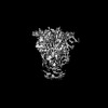
| Title | Cryo-EM density map of HKU1 spike glycoprotein D1 domain in complex with 9O-acetyl GD3 sialoglycan (Down_alt state, locally refined) | |||||||||
 Map data Map data | ||||||||||
 Sample Sample |
| |||||||||
 Keywords Keywords | HKU1 spike glycoprotein ectodomain / proline stablized / VIRAL PROTEIN | |||||||||
| Function / homology |  Function and homology information Function and homology informationhost cell endoplasmic reticulum-Golgi intermediate compartment membrane / receptor-mediated virion attachment to host cell / endocytosis involved in viral entry into host cell / fusion of virus membrane with host plasma membrane / fusion of virus membrane with host endosome membrane / viral envelope / host cell plasma membrane / virion membrane / membrane Similarity search - Function | |||||||||
| Biological species |  Human coronavirus HKU1 Human coronavirus HKU1 | |||||||||
| Method | single particle reconstruction / cryo EM / Resolution: 2.39 Å | |||||||||
 Authors Authors | Jin M / Rini JM | |||||||||
| Funding support |  Canada, 1 items Canada, 1 items
| |||||||||
 Citation Citation | Journal: Nat Commun / Year: 2025 Title: Human coronavirus HKU1 spike structures reveal the basis for sialoglycan specificity and carbohydrate-promoted conformational changes. Authors: Min Jin / Zaky Hassan / Zhijie Li / Ying Liu / Aleksandra Marakhovskaia / Alan H M Wong / Adam Forman / Mark Nitz / Michel Gilbert / Hai Yu / Xi Chen / James M Rini /  Abstract: The human coronavirus HKU1 uses both sialoglycoconjugates and the protein transmembrane serine protease 2 (TMPRSS2) as receptors. Carbohydrate binding leads to the spike protein up conformation ...The human coronavirus HKU1 uses both sialoglycoconjugates and the protein transmembrane serine protease 2 (TMPRSS2) as receptors. Carbohydrate binding leads to the spike protein up conformation required for TMPRSS2 binding, an outcome suggesting a distinct mechanism for driving fusion of the viral and host cell membranes. Nevertheless, the conformational changes promoted by carbohydrate binding have not been fully elucidated and the basis for HKU1's carbohydrate binding specificity remains unknown. Reported here are high resolution cryo-EM structures of the HKU1 spike protein trimer in its apo form and in complex with the carbohydrate moiety of a candidate carbohydrate receptor, the 9-O-acetylated GD3 ganglioside. The structures show that the spike monomer can exist in four discrete conformational states and that progression through them would promote the up conformation upon carbohydrate binding. We also show that a six-amino-acid insert is a determinant of HKU1's specificity for gangliosides containing a 9-O-acetylated α2-8-linked disialic acid moiety and that HKU1 shows weak affinity for the 9-O-acetylated sialic acids found on decoy receptors such as mucins. | |||||||||
| History |
|
- Structure visualization
| Supplemental images |
|---|

- Downloads & links
-EMDB archive
| Map data | emd_44885.map.gz | 83.9 MB | EMDB map data format | |
|---|---|---|---|---|
| Header (meta data) | emd-44885-v30.xmlemd-44885.xml | 18.8 KB 18.8 KB | Display Display | EMDB header |
| FSC (resolution estimation) | emd_44885_fsc.xml | 11.5 KB | Display | FSC data file |
| Images |  emd_44885.png emd_44885.png | 36.7 KB | ||
| Masks | emd_44885_msk_1.map | 166.4 MB | Mask map | |
| Filedesc metadata | emd-44885.cif.gz | 7.2 KB | ||
| Others | emd_44885_half_map_1.map.gzemd_44885_half_map_2.map.gz | 154.3 MB 154.3 MB | ||
| Archive directory |  http://ftp.pdbj.org/pub/emdb/structures/EMD-44885ftp://ftp.pdbj.org/pub/emdb/structures/EMD-44885 http://ftp.pdbj.org/pub/emdb/structures/EMD-44885ftp://ftp.pdbj.org/pub/emdb/structures/EMD-44885 | HTTPS FTP |
-Related structure data
| Related structure data |  9btaMC  9bswC  9bsxC  9bsyC  9bszC  9bt0C  9bt1C  9bt2C  9bt9C 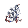 9btbC  9btcC  9btdC  9n12C  9n13C  9n14C  9n15C  9n16C  9n17C  9n18C  9n19C M: atomic model generated by this map C: citing same article ( |
|---|---|
| Similar structure data |


-Links
| EMDB pages | EMDB (EBI/PDBe) / EMDataResource |
|---|
-Map
| File | Download / File: emd_44885.map.gz / Format: CCP4 / Size: 166.4 MB / Type: IMAGE STORED AS FLOATING POINT NUMBER (4 BYTES) | ||||||||||||||||||||||||||||||||||||
|---|---|---|---|---|---|---|---|---|---|---|---|---|---|---|---|---|---|---|---|---|---|---|---|---|---|---|---|---|---|---|---|---|---|---|---|---|---|
| Projections & slices | Image control
Images are generated by Spider. | ||||||||||||||||||||||||||||||||||||
| Voxel size | X=Y=Z: 1.03 Å | ||||||||||||||||||||||||||||||||||||
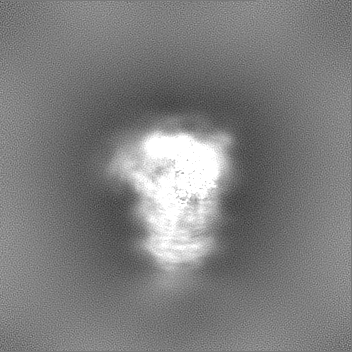
| Density |
| ||||||||||||||||||||||||||||||||||||
| Symmetry | Space group: 1 | ||||||||||||||||||||||||||||||||||||
| Details | EMDB XML:
|
 Z (Sec.)
 Y (Row.)
 X (Col.)


-Supplemental data
-Mask #1
| File | emd_44885_msk_1.map | ||||||||||||
|---|---|---|---|---|---|---|---|---|---|---|---|---|---|
| Projections & Slices |
| ||||||||||||
| Density Histograms |


-Half map: #1
| File | emd_44885_half_map_1.map | ||||||||||||
|---|---|---|---|---|---|---|---|---|---|---|---|---|---|
| Projections & Slices |
| ||||||||||||
| Density Histograms |


-Half map: #2
| File | emd_44885_half_map_2.map | ||||||||||||
|---|---|---|---|---|---|---|---|---|---|---|---|---|---|
| Projections & Slices |
| ||||||||||||
| Density Histograms |


- Sample components
-Entire : Human coronavirus HKU1 spike glycoprotein
| Entire | Name: Human coronavirus HKU1 spike glycoprotein |
|---|---|
| Components |
|
-Supramolecule #1: Human coronavirus HKU1 spike glycoprotein
| Supramolecule | Name: Human coronavirus HKU1 spike glycoprotein / type: complex / ID: 1 / Parent: 0 / Macromolecule list: #1 Details: Ectodomain generated by recombinant expression in HEK293 Freestyle cells |
|---|---|
| Source (natural) | Organism: Human coronavirus HKU1 |
| Molecular weight | Theoretical: 152.531 KDa |
-Macromolecule #1: Spike glycoprotein
| Macromolecule | Name: Spike glycoprotein / type: protein_or_peptide / ID: 1 / Number of copies: 1 / Enantiomer: LEVO |
|---|---|
| Source (natural) | Organism: Human coronavirus HKU1 |
| Molecular weight | Theoretical: 150.632734 KDa |
| Recombinant expression | Organism:  Homo sapiens (human) Homo sapiens (human) |
| Sequence | String: VIGDFNCTNF AINDLNTTVP RISEYVVDVS YGLGTYYILD RVYLNTTILF TGYFPKSGAN FRDLSLKGTT YLSTLWYQKP FLSDFNNGI FSRVKNTKLY VNKTLYSEFS TIVIGSVFIN NSYTIVVQPH NGVLEITACQ YTMCEYPHTI CKSKGSSRNE S WHFDKSEP ...String: VIGDFNCTNF AINDLNTTVP RISEYVVDVS YGLGTYYILD RVYLNTTILF TGYFPKSGAN FRDLSLKGTT YLSTLWYQKP FLSDFNNGI FSRVKNTKLY VNKTLYSEFS TIVIGSVFIN NSYTIVVQPH NGVLEITACQ YTMCEYPHTI CKSKGSSRNE S WHFDKSEP LCLFKKNFTY NVSTDWLYFH FYQERGTFYA YYADSGMPTT FLFSLYLGTL LSHYYVLPLT CNAISSNTDN ET LQYWVTP LSKRQYLLKF DNRGVITNAV DCSSSFFSEI QCKTKSLLPN TGVYDLSGFT VKPVATVHRR IPDLPDCDID KWL NNFNVP SPLNWERKIF SNCNFNLSTL LRLVHTDSFS CNNFDESKIY GSCFKSIVLD KFAIPNSRRS DLQLGSSGFL QSSN YKIDT TSSSCQLYYS LPAINVTINN YNPSSWNRRY GFNNFNLSSH SVVYSRYCFS VNNTFCPCAK PSFASSCKSH KPPSA SCPI GTNYRSCEST TVLDHTDWCR CSCLPDPITA YDPRSCSQKK SLVGVGEHCA GFGVDEEKCG VLDGSYNVSC LCSTDA FLG WSYDTCVSNN RCNIFSNFIL NGINSGTTCS NDLLQPNTEV FTDVCVDYDL YGITGQGIFK EVSAVYYNSW QNLLYDS NG NIIGFKDFVT NKTYNIFPCY AGRVSAAFHQ NASSLALLYR NLKCSYVLNN ISLTTQPYFD SYLGCVFNAD NLTDYSVS S CALRMGSGFC VDYNSPSSSS SRRKRRSISA SYRFVTFEPF NVSFVNDSIE SVGGLYEIKI PTNFTIVGQE EFIQTNSPK VTIDCSLFVC SNYAACHDLL SEYGTFCDNI NSILDEVNGL LDTTQLHVAD TLMQGVTLSS NLNTNLHFDV DNINFKSLVG CLGPHCGSS SRSFFEDLLF DKVKLSDVGF VEAYNNCTGG SEIRDLLCVQ SFNGIKVLPP ILSESQISGY TTAATVAAMF P PWSAAAGI PFSLNVQYRI NGLGVTMDVL NKNQKLIATA FNNALLSIQN GFSAPNSALA KIQSVVNSNA QALNSLLQQL FN KFGAISS SLQEILSRLD PPEAQVQIDR LINGRLTALN AYVSQQLSDI SLVKFGAALA MEKVNECVKS QSPRINFCGN GNH ILSLVQ NAPYGLLFMH FSYKPISFKT VLVSPGLCIS GDVGIAPKQG YFIKHNDHWM FTGSSYYYPE PISDKNVVFM NTCS VNFTK APLVYLNHSV PKLSDFESEL SHWFKNQTSI APNLTLNLHT INATFLDLYY EMNLIQESIK SLNNSYINLK DIGTY EMYV KSGGYIPEAP RDGQAYVRKD GEWVLLSTFL NSGRAHHHHH HGAGGLNDIF EAQKIEWHED TAAA UniProtKB: Spike glycoprotein |
-Macromolecule #5: 2-acetamido-2-deoxy-beta-D-glucopyranose
| Macromolecule | Name: 2-acetamido-2-deoxy-beta-D-glucopyranose / type: ligand / ID: 5 / Number of copies: 3 / Formula: NAG |
|---|---|
| Molecular weight | Theoretical: 221.208 Da |
| Chemical component information |  ChemComp-NAG: |
-Macromolecule #6: 5-acetamido-8-O-(5-acetamido-9-O-acetyl-3,5-dideoxy-D-glycero-alp...
| Macromolecule | Name: 5-acetamido-8-O-(5-acetamido-9-O-acetyl-3,5-dideoxy-D-glycero-alpha-D-galacto-non-2-ulopyranonosyl)-3,5-dideoxy-D-glycero-alpha-D-galacto-non-2-ulopyranosonic acid type: ligand / ID: 6 / Number of copies: 1 / Formula: A1AR1 |
|---|---|
| Molecular weight | Theoretical: 642.561 Da |
-Experimental details
-Structure determination
| Method | cryo EM |
|---|---|
 Processing Processing | single particle reconstruction |
| Aggregation state | particle |
-Sample preparation
| Concentration | 1.15 mg/mL | |||||||||
|---|---|---|---|---|---|---|---|---|---|---|
| Buffer | pH: 8 Component:
| |||||||||
| Grid | Model: C-flat-2/2 / Support film - Material: GOLD / Support film - topology: HOLEY / Support film - Film thickness: 20 | |||||||||
| Vitrification | Cryogen name: ETHANE / Chamber humidity: 100 % / Chamber temperature: 277 K / Instrument: FEI VITROBOT MARK IV |
- Electron microscopy
| Microscope | FEI TITAN KRIOS |
|---|---|
| Image recording | Film or detector model: FEI FALCON IV (4k x 4k) / Average electron dose: 36.0 e/Å2 |
| Electron beam | Acceleration voltage: 300 kV / Electron source:  FIELD EMISSION GUN FIELD EMISSION GUN |
| Electron optics | Illumination mode: FLOOD BEAM / Imaging mode: BRIGHT FIELD / Cs: 2.7 mm / Nominal defocus max: 1.8 µm / Nominal defocus min: 1.2 µm / Nominal magnification: 75000 |
| Sample stage | Specimen holder model: FEI TITAN KRIOS AUTOGRID HOLDER / Cooling holder cryogen: NITROGEN |
| Experimental equipment |  Model: Titan Krios / Image courtesy: FEI Company |
+Image processing

-Atomic model buiding 1
| Details | Symmetry-expanded particles are classified focused at D1 domain. Particles with D1 domain in active state are used in final local refinement and reconstruction with D1-only Mask. |
|---|---|
| Refinement | Protocol: FLEXIBLE FIT |
| Output model | PDB-9bta: |
