 Movie
Movie Controller

[English] 日本語
 Yorodumi
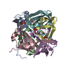
- PDB-9beo: Tungstate binding protein (Tungbindin) from Eubacterium limosum w... -
+ Open data

- Basic information
| Entry | Database: PDB / ID: 9beo | ||||||
|---|---|---|---|---|---|---|---|
| Title | Tungstate binding protein (Tungbindin) from Eubacterium limosum with 7.5 tungstates bound | ||||||
 Components Components | Molybdenum-pterin binding domain-containing protein | ||||||
 Keywords Keywords | METAL BINDING PROTEIN / Tungsten / metal binding / metal oxyanion / hexamer | ||||||
| Function / homology | Molybdenum-pterin binding domain / Mop domain profile. / Transport-associated OB, type 1 / TOBE domain / molybdate ion transport / Molybdate/tungstate binding, C-terminal / TUNGSTATE(VI)ION / Molybdenum-pterin-binding protein Function and homology information Function and homology information | ||||||
| Biological species |  Eubacterium limosum (bacteria) Eubacterium limosum (bacteria) | ||||||
| Method |  X-RAY DIFFRACTION / SAD / Resolution: 1.78 Å X-RAY DIFFRACTION / SAD / Resolution: 1.78 Å | ||||||
 Authors Authors | Zhou, D. / Rose, J.P. / Chen, L. / Wang, B.C. | ||||||
| Funding support |  United States, 1items United States, 1items
| ||||||
 Citation Citation | Journal: To Be Published Title: Tungstate binding protein (Tungbindin) from Eubacterium limosum with 7.5 tungstates bound Authors: Zhou, D. / Rose, J.P. / Chen, L. / Wang, B.C. | ||||||
| History |
|
- Structure visualization
| Structure viewer | Molecule: MolmilJmol/JSmol |
|---|
- Downloads & links
-Download
| PDBx/mmCIF format | 9beo.cif.gz | 98.5 KB | Display | PDBx/mmCIF format |
|---|---|---|---|---|
| PDB format | pdb9beo.ent.gz | 76 KB | Display | PDB format |
| PDBx/mmJSON format | 9beo.json.gz | Tree view | PDBx/mmJSON format | |
| Others |  Other downloads Other downloads |
-Validation report
| Arichive directory | https://data.pdbj.org/pub/pdb/validation_reports/be/9beoftp://data.pdbj.org/pub/pdb/validation_reports/be/9beo | HTTPS FTP |
|---|
-Related structure data
| Related structure data | |
|---|---|
| Similar structure data |


-Links
 PDBj
PDBj- Assembly
| Deposited unit | 
| ||||||||
|---|---|---|---|---|---|---|---|---|---|
| 1 |
| ||||||||
| Unit cell |
| ||||||||
| Components on special symmetry positions |
|
-Components
| #1: Protein | Mass: 8224.404 Da / Num. of mol.: 6 Source method: isolated from a genetically manipulated source Source: (gene. exp.) Eubacterium limosum (bacteria) / Gene: C7955_103236, SAMN04515624_10415 / Production host: #2: Chemical | ChemComp-WO4 /   Mass: 247.838 Da / Num. of mol.: 8 / Source method: obtained synthetically / Formula: WO4 / Feature type: SUBJECT OF INVESTIGATION Mass: 247.838 Da / Num. of mol.: 8 / Source method: obtained synthetically / Formula: WO4 / Feature type: SUBJECT OF INVESTIGATION#3: Water | ChemComp-HOH / |  Mass: 18.015 Da / Num. of mol.: 447 / Source method: isolated from a natural source / Formula: H2O Mass: 18.015 Da / Num. of mol.: 447 / Source method: isolated from a natural source / Formula: H2OHas ligand of interest | Y | |
|---|
-Experimental details
-Experiment
| Experiment | Method: X-RAY DIFFRACTION / Number of used crystals: 1 |
|---|
- Sample preparation
| Crystal | Density Matthews: 2.22 Å3/Da / Density % sol: 44.5 % |
|---|---|
| Crystal grow | Temperature: 293 K / Method: vapor diffusion, hanging drop Details: 0.2 M Sodium chloride, 0.1 M BIS-TRIS pH 6.5, 25% w/v Polyethylene glycol 3,350 |
-Data collection
| Diffraction | Mean temperature: 100 K / Serial crystal experiment: N |
|---|---|
| Diffraction source | Source: ROTATING ANODE / Type: RIGAKU MICROMAX-002 / Wavelength: 1.5418 Å |
| Detector | Type: DECTRIS PILATUS 200K / Detector: PIXEL / Date: Mar 19, 2024 |
| Radiation | Protocol: SINGLE WAVELENGTH / Monochromatic (M) / Laue (L): M / Scattering type: x-ray |
| Radiation wavelength | Wavelength: 1.5418 Å / Relative weight: 1 |
| Reflection | Resolution: 1.78→39.14 Å / Num. obs: 77439 / % possible obs: 96.5 % / Redundancy: 18 % / Rmerge(I) obs: 0.097 / Rpim(I) all: 0.022 / Rrim(I) all: 0.1 / Net I/σ(I): 52.7 |
| Reflection shell | Resolution: 1.78→1.81 Å / Redundancy: 5 % / Rmerge(I) obs: 0.446 / Mean I/σ(I) obs: 3.76 / Num. unique obs: 1071 / CC1/2: 0.86 / CC star: 0.962 / Rpim(I) all: 0.21 / Rrim(I) all: 0.497 / % possible all: 70.7 |
- Processing
| Software |
| ||||||||||||||||||||||||||||||||||||||||||||||||||||||||||||||||||||||||||||||||||||||||||||||||||||||||||||||||||||||||||||||||||||||||||||||||||||||||||||||||||||||||||||||||||||||||||||||||||||
|---|---|---|---|---|---|---|---|---|---|---|---|---|---|---|---|---|---|---|---|---|---|---|---|---|---|---|---|---|---|---|---|---|---|---|---|---|---|---|---|---|---|---|---|---|---|---|---|---|---|---|---|---|---|---|---|---|---|---|---|---|---|---|---|---|---|---|---|---|---|---|---|---|---|---|---|---|---|---|---|---|---|---|---|---|---|---|---|---|---|---|---|---|---|---|---|---|---|---|---|---|---|---|---|---|---|---|---|---|---|---|---|---|---|---|---|---|---|---|---|---|---|---|---|---|---|---|---|---|---|---|---|---|---|---|---|---|---|---|---|---|---|---|---|---|---|---|---|---|---|---|---|---|---|---|---|---|---|---|---|---|---|---|---|---|---|---|---|---|---|---|---|---|---|---|---|---|---|---|---|---|---|---|---|---|---|---|---|---|---|---|---|---|---|---|---|---|---|
| Refinement | Method to determine structure: SAD / Resolution: 1.78→39.14 Å / SU ML: 0.16 / Cross valid method: FREE R-VALUE / σ(F): 1.33 / Phase error: 20.16 / Stereochemistry target values: ML
| ||||||||||||||||||||||||||||||||||||||||||||||||||||||||||||||||||||||||||||||||||||||||||||||||||||||||||||||||||||||||||||||||||||||||||||||||||||||||||||||||||||||||||||||||||||||||||||||||||||
| Solvent computation | Shrinkage radii: 0.9 Å / VDW probe radii: 1.1 Å / Solvent model: FLAT BULK SOLVENT MODEL | ||||||||||||||||||||||||||||||||||||||||||||||||||||||||||||||||||||||||||||||||||||||||||||||||||||||||||||||||||||||||||||||||||||||||||||||||||||||||||||||||||||||||||||||||||||||||||||||||||||
| Refinement step | Cycle: LAST / Resolution: 1.78→39.14 Å
| ||||||||||||||||||||||||||||||||||||||||||||||||||||||||||||||||||||||||||||||||||||||||||||||||||||||||||||||||||||||||||||||||||||||||||||||||||||||||||||||||||||||||||||||||||||||||||||||||||||
| Refine LS restraints |
| ||||||||||||||||||||||||||||||||||||||||||||||||||||||||||||||||||||||||||||||||||||||||||||||||||||||||||||||||||||||||||||||||||||||||||||||||||||||||||||||||||||||||||||||||||||||||||||||||||||
| LS refinement shell |
|
