 Movie
Movie Controller


+ Open data

- Basic information
| Entry | Database: PDB / ID: 9bdv | ||||||
|---|---|---|---|---|---|---|---|
| Title | NF-kappaB RelA homo-dimer bound to TA-centric kappaB DNA | ||||||
 Components Components |
| ||||||
 Keywords Keywords | DNA BINDING PROTEIN/DNA / RelA / kappaB DNA / Promoter / Transcription / DNA BINDING PROTEIN / DNA BINDING PROTEIN-DNA complex | ||||||
| Function / homology |  Function and homology information Function and homology informationSUMOylation of immune response proteins / Regulated proteolysis of p75NTR / DEx/H-box helicases activate type I IFN and inflammatory cytokines production / Interleukin-1 processing / RIP-mediated NFkB activation via ZBP1 / TRAF6 mediated NF-kB activation / positive regulation of Schwann cell differentiation / PKMTs methylate histone lysines / NF-kB is activated and signals survival / Turbulent (oscillatory, disturbed) flow shear stress activates signaling by PIEZO1 and integrins in endothelial cells ...SUMOylation of immune response proteins / Regulated proteolysis of p75NTR / DEx/H-box helicases activate type I IFN and inflammatory cytokines production / Interleukin-1 processing / RIP-mediated NFkB activation via ZBP1 / TRAF6 mediated NF-kB activation / positive regulation of Schwann cell differentiation / PKMTs methylate histone lysines / NF-kB is activated and signals survival / Turbulent (oscillatory, disturbed) flow shear stress activates signaling by PIEZO1 and integrins in endothelial cells / TAK1-dependent IKK and NF-kappa-B activation / Activation of NF-kappaB in B cells / prolactin signaling pathway / positive regulation of chondrocyte differentiation / NF-kappaB p50/p65 complex / toll-like receptor TLR6:TLR2 signaling pathway / CLEC7A (Dectin-1) signaling / FCERI mediated NF-kB activation / Interleukin-1 signaling / Downstream TCR signaling / CD209 (DC-SIGN) signaling / cellular response to peptidoglycan / ankyrin repeat binding / nucleotide-binding oligomerization domain containing 2 signaling pathway / negative regulation of protein sumoylation / postsynapse to nucleus signaling pathway / defense response to tumor cell / positive regulation of T cell receptor signaling pathway / signal transduction involved in regulation of gene expression / cellular response to interleukin-6 / negative regulation of non-canonical NF-kappaB signal transduction / actinin binding / positive regulation of miRNA metabolic process / response to UV-B / interleukin-1-mediated signaling pathway / positive regulation of leukocyte adhesion to vascular endothelial cell / toll-like receptor 4 signaling pathway / cellular response to hepatocyte growth factor stimulus / positive regulation of amyloid-beta formation / vascular endothelial growth factor signaling pathway / non-canonical NF-kappaB signal transduction / NF-kappaB complex / phosphate ion binding / cellular response to lipoteichoic acid / response to muramyl dipeptide / cellular response to angiotensin / hair follicle development / positive regulation of vascular endothelial growth factor production / cellular response to interleukin-1 / general transcription initiation factor binding / NF-kappaB binding / RNA polymerase II core promoter sequence-specific DNA binding / canonical NF-kappaB signal transduction / response to muscle stretch / positive regulation of interleukin-12 production / peptide binding / negative regulation of insulin receptor signaling pathway / negative regulation of cytokine production involved in inflammatory response / response to cytokine / negative regulation of miRNA transcription / negative regulation of angiogenesis / response to interleukin-1 / antiviral innate immune response / : / animal organ morphogenesis / tumor necrosis factor-mediated signaling pathway / positive regulation of interleukin-8 production / positive regulation of interleukin-1 beta production / negative regulation of extrinsic apoptotic signaling pathway / response to bacterium / RNA polymerase II transcription regulatory region sequence-specific DNA binding / liver development / defense response / neuropeptide signaling pathway / negative regulation of protein catabolic process / chromatin DNA binding / positive regulation of non-canonical NF-kappaB signal transduction / cellular response to nicotine / positive regulation of miRNA transcription / positive regulation of interleukin-6 production / DNA-binding transcription repressor activity, RNA polymerase II-specific / cellular response to tumor necrosis factor / transcription coactivator binding / histone deacetylase binding / cellular response to hydrogen peroxide / cytokine-mediated signaling pathway / chromatin organization / cellular response to lipopolysaccharide / regulation of inflammatory response / double-stranded DNA binding / DNA-binding transcription activator activity, RNA polymerase II-specific / transcription regulator complex / sequence-specific DNA binding / DNA-binding transcription factor binding / DNA-binding transcription factor activity, RNA polymerase II-specific / positive regulation of canonical NF-kappaB signal transduction / transcription coactivator activity / transcription cis-regulatory region binding / intracellular signal transduction / RNA polymerase II cis-regulatory region sequence-specific DNA binding Similarity search - Function | ||||||
| Biological species |   Homo sapiens (human) Homo sapiens (human) | ||||||
| Method |  X-RAY DIFFRACTION / SYNCHROTRON / MOLECULAR REPLACEMENT / Resolution: 1.9 Å X-RAY DIFFRACTION / SYNCHROTRON / MOLECULAR REPLACEMENT / Resolution: 1.9 Å | ||||||
 Authors Authors | Biswas, T. / Shahabi, S. / Tsodikov, O.V. | ||||||
| Funding support |  United States, 1items United States, 1items
| ||||||
 Citation Citation | Journal: Proc.Natl.Acad.Sci.USA / Year: 2024 Title: Transient interactions modulate the affinity of NF-kappa B transcription factors for DNA. Authors: Li, T. / Shahabi, S. / Biswas, T. / Tsodikov, O.V. / Pan, W. / Huang, D.B. / Wang, V.Y. / Wang, Y. / Ghosh, G. | ||||||
| History |
|
- Structure visualization
| Structure viewer | Molecule: MolmilJmol/JSmol |
|---|
- Downloads & links
-Download
| PDBx/mmCIF format | 9bdv.cif.gz | 298.5 KB | Display | PDBx/mmCIF format |
|---|---|---|---|---|
| PDB format | pdb9bdv.ent.gz | 235.8 KB | Display | PDB format |
| PDBx/mmJSON format | 9bdv.json.gz | Tree view | PDBx/mmJSON format | |
| Others |  Other downloads Other downloads |
-Validation report
| Arichive directory | https://data.pdbj.org/pub/pdb/validation_reports/bd/9bdvftp://data.pdbj.org/pub/pdb/validation_reports/bd/9bdv | HTTPS FTP |
|---|
-Related structure data
| Related structure data | 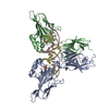 9bduC 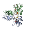 9bdwC  9bdxC 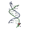 9bdyC  9bdzC  9be0C  9be1C 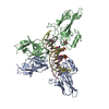 2ramS S: Starting model for refinement C: citing same article ( |
|---|---|
| Similar structure data |
-Links
 PDBj
PDBj


- Assembly
| Deposited unit | 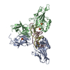
| ||||||||
|---|---|---|---|---|---|---|---|---|---|
| 1 |
| ||||||||
| Unit cell |
|
-Components
| #1: Protein | Mass: 32869.223 Da / Num. of mol.: 2 / Fragment: residues 19-304 Source method: isolated from a genetically manipulated source Source: (gene. exp.)  #2: DNA chain | | Mass: 5859.809 Da / Num. of mol.: 1 / Source method: obtained synthetically / Source: (synth.) Homo sapiens (human)#3: DNA chain | | Mass: 5788.773 Da / Num. of mol.: 1 / Source method: obtained synthetically / Source: (synth.) Homo sapiens (human)#4: Chemical | ChemComp-SO4 /   Mass: 96.063 Da / Num. of mol.: 4 / Source method: obtained synthetically / Formula: SO4 / Feature type: SUBJECT OF INVESTIGATION Mass: 96.063 Da / Num. of mol.: 4 / Source method: obtained synthetically / Formula: SO4 / Feature type: SUBJECT OF INVESTIGATION#5: Water | ChemComp-HOH / |  Mass: 18.015 Da / Num. of mol.: 603 / Source method: isolated from a natural source / Formula: H2O Mass: 18.015 Da / Num. of mol.: 603 / Source method: isolated from a natural source / Formula: H2OHas ligand of interest | Y | Has protein modification | N | |
|---|
-Experimental details
-Experiment
| Experiment | Method: X-RAY DIFFRACTION / Number of used crystals: 1 |
|---|
- Sample preparation
| Crystal | Density Matthews: 2.32 Å3/Da / Density % sol: 47.04 % |
|---|---|
| Crystal grow | Temperature: 293 K / Method: vapor diffusion, hanging drop / Details: PEG3350, MES, Spermine, beta-octylglucoside |
-Data collection
| Diffraction | Mean temperature: 80 K / Serial crystal experiment: N | ||||||||||||||||||||||||||||||||||||||||||||||||||||||||||||||||||||||||||||||||||||||||||||||||||||||||||||||||||||||||||||||||||||||||||||
|---|---|---|---|---|---|---|---|---|---|---|---|---|---|---|---|---|---|---|---|---|---|---|---|---|---|---|---|---|---|---|---|---|---|---|---|---|---|---|---|---|---|---|---|---|---|---|---|---|---|---|---|---|---|---|---|---|---|---|---|---|---|---|---|---|---|---|---|---|---|---|---|---|---|---|---|---|---|---|---|---|---|---|---|---|---|---|---|---|---|---|---|---|---|---|---|---|---|---|---|---|---|---|---|---|---|---|---|---|---|---|---|---|---|---|---|---|---|---|---|---|---|---|---|---|---|---|---|---|---|---|---|---|---|---|---|---|---|---|---|---|---|
| Diffraction source | Source: SYNCHROTRON / Site: APS / Beamline: 19-ID / Wavelength: 0.9793 Å | ||||||||||||||||||||||||||||||||||||||||||||||||||||||||||||||||||||||||||||||||||||||||||||||||||||||||||||||||||||||||||||||||||||||||||||
| Detector | Type: ADSC QUANTUM 315 / Detector: CCD / Date: Nov 19, 2013 | ||||||||||||||||||||||||||||||||||||||||||||||||||||||||||||||||||||||||||||||||||||||||||||||||||||||||||||||||||||||||||||||||||||||||||||
| Radiation | Protocol: SINGLE WAVELENGTH / Monochromatic (M) / Laue (L): M / Scattering type: x-ray | ||||||||||||||||||||||||||||||||||||||||||||||||||||||||||||||||||||||||||||||||||||||||||||||||||||||||||||||||||||||||||||||||||||||||||||
| Radiation wavelength | Wavelength: 0.9793 Å / Relative weight: 1 | ||||||||||||||||||||||||||||||||||||||||||||||||||||||||||||||||||||||||||||||||||||||||||||||||||||||||||||||||||||||||||||||||||||||||||||
| Reflection | Resolution: 1.9→30 Å / Num. obs: 57610 / % possible obs: 99.6 % / Redundancy: 7.8 % / Rmerge(I) obs: 0.994 / Χ2: 0.047 / Net I/σ(I): 8.5 / Num. measured all: 448525 | ||||||||||||||||||||||||||||||||||||||||||||||||||||||||||||||||||||||||||||||||||||||||||||||||||||||||||||||||||||||||||||||||||||||||||||
| Reflection shell |
|
- Processing
| Software |
| ||||||||||||||||||||||||||||||||||||||||||||||||||||||||||||||||||||||||||||||||||||||||||||||||||||||||||||||||||||||||||||||||||||||||||||||||||||||||||||||||||||||||||||||||||||||
|---|---|---|---|---|---|---|---|---|---|---|---|---|---|---|---|---|---|---|---|---|---|---|---|---|---|---|---|---|---|---|---|---|---|---|---|---|---|---|---|---|---|---|---|---|---|---|---|---|---|---|---|---|---|---|---|---|---|---|---|---|---|---|---|---|---|---|---|---|---|---|---|---|---|---|---|---|---|---|---|---|---|---|---|---|---|---|---|---|---|---|---|---|---|---|---|---|---|---|---|---|---|---|---|---|---|---|---|---|---|---|---|---|---|---|---|---|---|---|---|---|---|---|---|---|---|---|---|---|---|---|---|---|---|---|---|---|---|---|---|---|---|---|---|---|---|---|---|---|---|---|---|---|---|---|---|---|---|---|---|---|---|---|---|---|---|---|---|---|---|---|---|---|---|---|---|---|---|---|---|---|---|---|---|
| Refinement | Method to determine structure: MOLECULAR REPLACEMENT Starting model: 2RAM Resolution: 1.9→29.66 Å / Cor.coef. Fo:Fc: 0.947 / Cor.coef. Fo:Fc free: 0.925 / SU B: 8.212 / SU ML: 0.105 / Cross valid method: THROUGHOUT / ESU R Free: 0.156 / Stereochemistry target values: MAXIMUM LIKELIHOOD / Details: HYDROGENS HAVE BEEN ADDED IN THE RIDING POSITIONS
| ||||||||||||||||||||||||||||||||||||||||||||||||||||||||||||||||||||||||||||||||||||||||||||||||||||||||||||||||||||||||||||||||||||||||||||||||||||||||||||||||||||||||||||||||||||||
| Solvent computation | Ion probe radii: 0.8 Å / Shrinkage radii: 0.8 Å / VDW probe radii: 1.2 Å / Solvent model: MASK | ||||||||||||||||||||||||||||||||||||||||||||||||||||||||||||||||||||||||||||||||||||||||||||||||||||||||||||||||||||||||||||||||||||||||||||||||||||||||||||||||||||||||||||||||||||||
| Displacement parameters | Biso mean: 26.612 Å2
| ||||||||||||||||||||||||||||||||||||||||||||||||||||||||||||||||||||||||||||||||||||||||||||||||||||||||||||||||||||||||||||||||||||||||||||||||||||||||||||||||||||||||||||||||||||||
| Refinement step | Cycle: 1 / Resolution: 1.9→29.66 Å
| ||||||||||||||||||||||||||||||||||||||||||||||||||||||||||||||||||||||||||||||||||||||||||||||||||||||||||||||||||||||||||||||||||||||||||||||||||||||||||||||||||||||||||||||||||||||
| Refine LS restraints |
|