 Movie
Movie Controller

[English] 日本語
 Yorodumi
- PDB-9bbl: THF filament generated from 4E-Tau(297-407) under neutral Mg2+ co... -
+ Open data

- Basic information
| Entry | Database: PDB / ID: 9bbl | ||||||
|---|---|---|---|---|---|---|---|
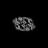
| Title | THF filament generated from 4E-Tau(297-407) under neutral Mg2+ condition | ||||||
 Components Components | Isoform Tau-F of Microtubule-associated protein tau | ||||||
 Keywords Keywords | PROTEIN FIBRIL / Tau / amyloid fibrils / in vitro / PHF | ||||||
| Function / homology |  Function and homology information Function and homology informationplus-end-directed organelle transport along microtubule / histone-dependent DNA binding / negative regulation of establishment of protein localization to mitochondrion / neurofibrillary tangle / microtubule lateral binding / axonal transport / tubulin complex / positive regulation of protein localization to synapse / negative regulation of tubulin deacetylation / phosphatidylinositol bisphosphate binding ...plus-end-directed organelle transport along microtubule / histone-dependent DNA binding / negative regulation of establishment of protein localization to mitochondrion / neurofibrillary tangle / microtubule lateral binding / axonal transport / tubulin complex / positive regulation of protein localization to synapse / negative regulation of tubulin deacetylation / phosphatidylinositol bisphosphate binding / generation of neurons / rRNA metabolic process / axonal transport of mitochondrion / regulation of mitochondrial fission / axon development / regulation of chromosome organization / central nervous system neuron development / intracellular distribution of mitochondria / minor groove of adenine-thymine-rich DNA binding / lipoprotein particle binding / microtubule polymerization / negative regulation of mitochondrial membrane potential / dynactin binding / regulation of microtubule polymerization / apolipoprotein binding / main axon / protein polymerization / axolemma / glial cell projection / Caspase-mediated cleavage of cytoskeletal proteins / regulation of microtubule polymerization or depolymerization / negative regulation of mitochondrial fission / neurofibrillary tangle assembly / positive regulation of axon extension / regulation of cellular response to heat / Activation of AMPK downstream of NMDARs / synapse assembly / positive regulation of superoxide anion generation / regulation of long-term synaptic depression / positive regulation of protein localization / supramolecular fiber organization / cellular response to brain-derived neurotrophic factor stimulus / regulation of calcium-mediated signaling / cytoplasmic microtubule organization / positive regulation of microtubule polymerization / somatodendritic compartment / axon cytoplasm / astrocyte activation / stress granule assembly / phosphatidylinositol binding / nuclear periphery / regulation of microtubule cytoskeleton organization / protein phosphatase 2A binding / cellular response to reactive oxygen species / Hsp90 protein binding / microglial cell activation / cellular response to nerve growth factor stimulus / synapse organization / protein homooligomerization / PKR-mediated signaling / regulation of synaptic plasticity / SH3 domain binding / response to lead ion / microtubule cytoskeleton organization / memory / cytoplasmic ribonucleoprotein granule / neuron projection development / cell-cell signaling / single-stranded DNA binding / protein-folding chaperone binding / cellular response to heat / actin binding / microtubule cytoskeleton / cell body / growth cone / double-stranded DNA binding / protein-macromolecule adaptor activity / microtubule binding / dendritic spine / sequence-specific DNA binding / amyloid fibril formation / microtubule / learning or memory / neuron projection / regulation of autophagy / membrane raft / axon / negative regulation of gene expression / neuronal cell body / dendrite / DNA damage response / protein kinase binding / enzyme binding / mitochondrion / DNA binding / RNA binding / extracellular region / identical protein binding / nucleus / plasma membrane Similarity search - Function | ||||||
| Biological species |  Homo sapiens (human) Homo sapiens (human) | ||||||
| Method | ELECTRON MICROSCOPY / helical reconstruction / cryo EM / Resolution: 2.5 Å | ||||||
 Authors Authors | Duan, P. / El Mammeri, N. | ||||||
| Funding support |  United States, 1items United States, 1items
| ||||||
 Citation Citation | Journal: J Biol Chem / Year: 2024 Title: Milligram-scale assembly and NMR fingerprint of tau fibrils adopting the Alzheimer's disease fold. Authors: Pu Duan / Nadia El Mammeri / Mei Hong / Abstract: In the Alzheimer's disease (AD) brain, the microtubule-associated protein tau aggregates into paired helical filaments in which each protofilament has a C-shaped conformation. In vitro assembly of ...In the Alzheimer's disease (AD) brain, the microtubule-associated protein tau aggregates into paired helical filaments in which each protofilament has a C-shaped conformation. In vitro assembly of tau fibrils adopting this fold is highly valuable for both fundamental and applied studies of AD without requiring patient-brain extracted fibrils. To date, reported methods for forming AD-fold tau fibrils have been irreproducible and sensitive to subtle variations in fibrillization conditions. Here, we describe a route to reproducibly assemble tau fibrils adopting the AD fold on the multi-milligram scale. We investigated the fibrillization conditions of two constructs and found that a tau (297-407) construct that contains four AD phospho-mimetic glutamate mutations robustly formed the C-shaped conformation. 2D and 3D correlation solid-state NMR spectra show a single predominant set of chemical shifts, indicating a single molecular conformation. Negative-stain electron microscopy and cryo-EM data confirm that the protofilament formed by 4E-tau (297-407) adopts the C-shaped conformation, which associates into paired, triple, and quadruple helical filaments. In comparison, NMR spectra indicate that a previously reported construct, tau (297-391), forms a mixture of a four-layered dimer structure and the C-shaped structure, whose populations are sensitive to the environmental conditions. The determination of the NMR chemical shifts of the AD-fold tau opens the possibility for future studies of tau fibril conformations and ligand binding by NMR. The quantitative assembly of tau fibrils adopting the AD fold should facilitate the development of diagnostic and therapeutic compounds that target AD tau. | ||||||
| History |
|
- Structure visualization
| Structure viewer | Molecule: MolmilJmol/JSmol |
|---|
- Downloads & links
-Download
| PDBx/mmCIF format | 9bbl.cif.gz | 260.9 KB | Display | PDBx/mmCIF format |
|---|---|---|---|---|
| PDB format | pdb9bbl.ent.gz | 189.5 KB | Display | PDB format |
| PDBx/mmJSON format | 9bbl.json.gz | Tree view | PDBx/mmJSON format | |
| Others |  Other downloads Other downloads |
-Validation report
| Summary document | 9bbl_validation.pdf.gz | 1.2 MB | Display | wwPDB validaton report |
|---|---|---|---|---|
| Full document | 9bbl_full_validation.pdf.gz | 1.2 MB | Display | |
| Data in XML | 9bbl_validation.xml.gz | 37 KB | Display | |
| Data in CIF | 9bbl_validation.cif.gz | 54.4 KB | Display | |
| Arichive directory | https://data.pdbj.org/pub/pdb/validation_reports/bb/9bblftp://data.pdbj.org/pub/pdb/validation_reports/bb/9bbl | HTTPS FTP |
-Related structure data
| Related structure data | 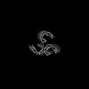 44421MC 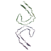 9bbmC C: citing same article ( M: map data used to model this data |
|---|---|
| Similar structure data |

-Links
 PDBj
PDBj


- Assembly
| Deposited unit | 
|
|---|---|
| 1 |
|
-Components
| #1: Protein | Mass: 46073.988 Da / Num. of mol.: 9 / Mutation: S396E, S400E, T403E, S404E Source method: isolated from a genetically manipulated source Source: (gene. exp.) Homo sapiens (human) / Gene: MAPT, MAPTL, MTBT1, TAU / Production host:  |
|---|
-Experimental details
-Experiment
| Experiment | Method: ELECTRON MICROSCOPY |
|---|---|
| EM experiment | Aggregation state: FILAMENT / 3D reconstruction method: helical reconstruction |
- Sample preparation
| Component | Name: In vitro 4E-tau (297-407) THF fibrils / Type: COMPLEX / Entity ID: all / Source: RECOMBINANT | ||||||||||||||||||||
|---|---|---|---|---|---|---|---|---|---|---|---|---|---|---|---|---|---|---|---|---|---|
| Molecular weight | Experimental value: NO | ||||||||||||||||||||
| Source (natural) | Organism: Homo sapiens (human) | ||||||||||||||||||||
| Source (recombinant) | Organism: | ||||||||||||||||||||
| Buffer solution | pH: 7.4 Details: The pH was tuned to pH 7.5-8 using 5M NaOH after mixing phosphate buffer and MgCl2, and precipitation was formed. | ||||||||||||||||||||
| Buffer component |
| ||||||||||||||||||||
| Specimen | Conc.: 6 mg/ml / Embedding applied: NO / Shadowing applied: NO / Staining applied: NO / Vitrification applied: YES | ||||||||||||||||||||
| Specimen support | Grid material: COPPER / Grid mesh size: 300 divisions/in. / Grid type: Quantifoil R1.2/1.3 | ||||||||||||||||||||
| Vitrification | Instrument: FEI VITROBOT MARK IV / Cryogen name: ETHANE / Humidity: 95 % / Chamber temperature: 277 K |
- Electron microscopy imaging
| Experimental equipment |  Model: Titan Krios / Image courtesy: FEI Company |
|---|---|
| Microscopy | Model: TFS KRIOS |
| Electron gun | Electron source:  FIELD EMISSION GUN / Accelerating voltage: 300 kV / Illumination mode: SPOT SCAN FIELD EMISSION GUN / Accelerating voltage: 300 kV / Illumination mode: SPOT SCAN |
| Electron lens | Mode: BRIGHT FIELD / Nominal defocus max: 2000 nm / Nominal defocus min: 200 nm |
| Image recording | Electron dose: 31.004 e/Å2 / Film or detector model: GATAN K3 BIOCONTINUUM (6k x 4k) |
- Processing
| EM software |
| ||||||||||||||||||||
|---|---|---|---|---|---|---|---|---|---|---|---|---|---|---|---|---|---|---|---|---|---|
| CTF correction | Type: PHASE FLIPPING AND AMPLITUDE CORRECTION | ||||||||||||||||||||
| Helical symmerty | Angular rotation/subunit: -0.841 ° / Axial rise/subunit: 4.745 Å / Axial symmetry: C1 | ||||||||||||||||||||
| Particle selection | Num. of particles selected: 651965 / Details: Manual Selection of Start-End Coordinates | ||||||||||||||||||||
| 3D reconstruction | Resolution: 2.5 Å / Resolution method: FSC 0.143 CUT-OFF / Num. of particles: 56126 / Symmetry type: HELICAL |
