 Movie
Movie Controller


+ Open data

- Basic information
| Entry | 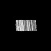 | |||||||||
|---|---|---|---|---|---|---|---|---|---|---|
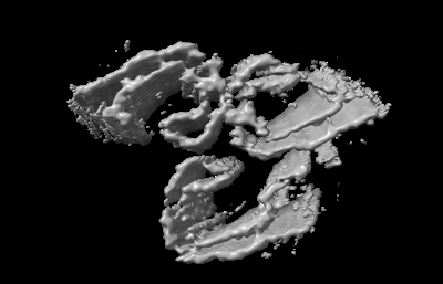
| Title | Tau(291-407)-4E THF | |||||||||
 Map data Map data | ||||||||||
 Sample Sample |
| |||||||||
 Keywords Keywords | Tau / amyloid fibrils / PHF / QHF / STRUCTURAL PROTEIN | |||||||||
| Biological species |  | |||||||||
| Method | helical reconstruction / cryo EM / Resolution: 4.5 Å | |||||||||
 Authors Authors | El Mammeri N / Duan P / Hong M | |||||||||
| Funding support |  United States, 1 items United States, 1 items
| |||||||||
 Citation Citation | Journal: J Biol Chem / Year: 2024 Title: Milligram-scale assembly and NMR fingerprint of tau fibrils adopting the Alzheimer's disease fold. Authors: Pu Duan / Nadia El Mammeri / Mei Hong / Abstract: In the Alzheimer's disease (AD) brain, the microtubule-associated protein tau aggregates into paired helical filaments in which each protofilament has a C-shaped conformation. In vitro assembly of ...In the Alzheimer's disease (AD) brain, the microtubule-associated protein tau aggregates into paired helical filaments in which each protofilament has a C-shaped conformation. In vitro assembly of tau fibrils adopting this fold is highly valuable for both fundamental and applied studies of AD without requiring patient-brain extracted fibrils. To date, reported methods for forming AD-fold tau fibrils have been irreproducible and sensitive to subtle variations in fibrillization conditions. Here, we describe a route to reproducibly assemble tau fibrils adopting the AD fold on the multi-milligram scale. We investigated the fibrillization conditions of two constructs and found that a tau (297-407) construct that contains four AD phospho-mimetic glutamate mutations robustly formed the C-shaped conformation. 2D and 3D correlation solid-state NMR spectra show a single predominant set of chemical shifts, indicating a single molecular conformation. Negative-stain electron microscopy and cryo-EM data confirm that the protofilament formed by 4E-tau (297-407) adopts the C-shaped conformation, which associates into paired, triple, and quadruple helical filaments. In comparison, NMR spectra indicate that a previously reported construct, tau (297-391), forms a mixture of a four-layered dimer structure and the C-shaped structure, whose populations are sensitive to the environmental conditions. The determination of the NMR chemical shifts of the AD-fold tau opens the possibility for future studies of tau fibril conformations and ligand binding by NMR. The quantitative assembly of tau fibrils adopting the AD fold should facilitate the development of diagnostic and therapeutic compounds that target AD tau. | |||||||||
| History |
|
- Structure visualization
| Supplemental images |
|---|
- Downloads & links
-EMDB archive
| Map data | emd_43826.map.gz | 10.8 MB |  EMDB map data format EMDB map data format | |
|---|---|---|---|---|
| Header (meta data) | emd-43826-v30.xmlemd-43826.xml | 14.1 KB 14.1 KB | Display Display | EMDB header |
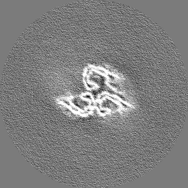
| Images |  emd_43826.png emd_43826.png | 48.8 KB | ||
| Masks | emd_43826_msk_1.map | 216 MB | Mask map | |
| Filedesc metadata | emd-43826.cif.gz | 3.8 KB | ||
| Others | emd_43826_additional_1.map.gzemd_43826_half_map_1.map.gzemd_43826_half_map_2.map.gz | 170.9 MB 171.2 MB 171.2 MB | ||
| Archive directory |  http://ftp.pdbj.org/pub/emdb/structures/EMD-43826ftp://ftp.pdbj.org/pub/emdb/structures/EMD-43826 http://ftp.pdbj.org/pub/emdb/structures/EMD-43826ftp://ftp.pdbj.org/pub/emdb/structures/EMD-43826 | HTTPS FTP |
-Validation report
| Summary document | emd_43826_validation.pdf.gz | 886.5 KB | Display | EMDB validaton report |
|---|---|---|---|---|
| Full document | emd_43826_full_validation.pdf.gz | 886 KB | Display | |
| Data in XML | emd_43826_validation.xml.gz | 15.6 KB | Display | |
| Data in CIF | emd_43826_validation.cif.gz | 18.7 KB | Display | |
| Arichive directory | https://ftp.pdbj.org/pub/emdb/validation_reports/EMD-43826ftp://ftp.pdbj.org/pub/emdb/validation_reports/EMD-43826 | HTTPS FTP |
-Related structure data

-Links
| EMDB pages | EMDB (EBI/PDBe) / EMDataResource |
|---|
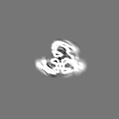
-Map
| File | Download / File: emd_43826.map.gz / Format: CCP4 / Size: 216 MB / Type: IMAGE STORED AS FLOATING POINT NUMBER (4 BYTES) | ||||||||||||||||||||||||||||||||||||
|---|---|---|---|---|---|---|---|---|---|---|---|---|---|---|---|---|---|---|---|---|---|---|---|---|---|---|---|---|---|---|---|---|---|---|---|---|---|
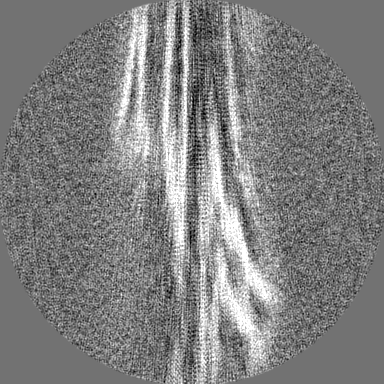
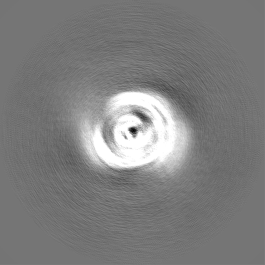
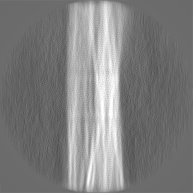
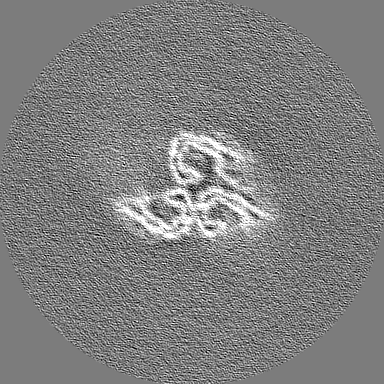
| Projections & slices | Image control
Images are generated by Spider. | ||||||||||||||||||||||||||||||||||||
| Voxel size | X=Y=Z: 1.06 Å | ||||||||||||||||||||||||||||||||||||
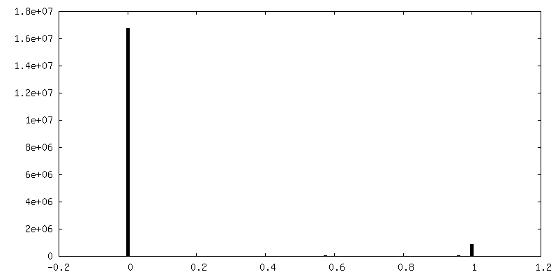
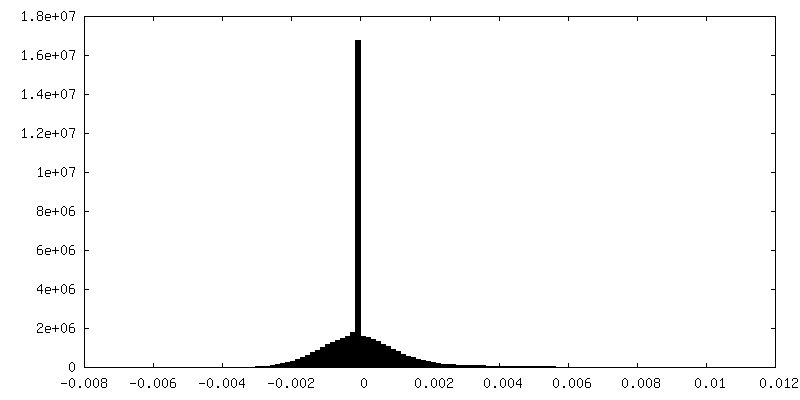
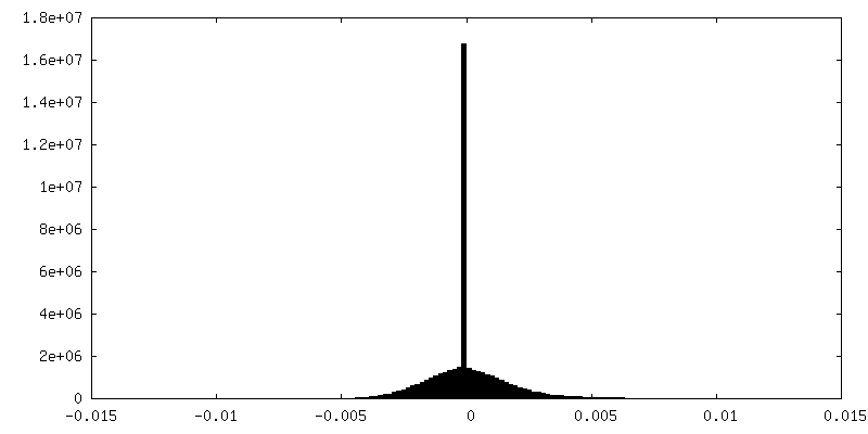
| Density |
| ||||||||||||||||||||||||||||||||||||
| Symmetry | Space group: 1 | ||||||||||||||||||||||||||||||||||||
| Details | EMDB XML:
|
 Z (Sec.)
 Y (Row.)
 X (Col.)


-Supplemental data
-Mask #1
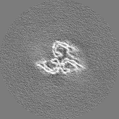
| File | emd_43826_msk_1.map | ||||||||||||
|---|---|---|---|---|---|---|---|---|---|---|---|---|---|
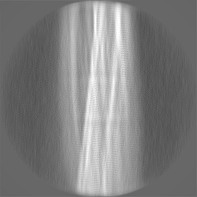
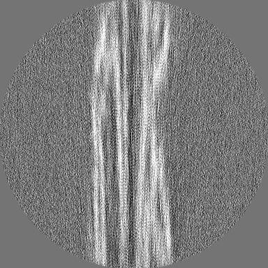
| Projections & Slices |
| ||||||||||||
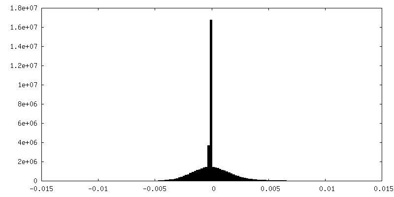
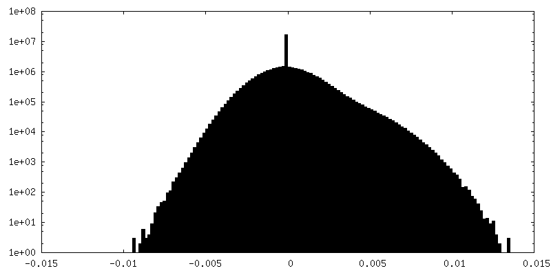
| Density Histograms |


-Additional map: #1
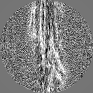
| File | emd_43826_additional_1.map | ||||||||||||
|---|---|---|---|---|---|---|---|---|---|---|---|---|---|
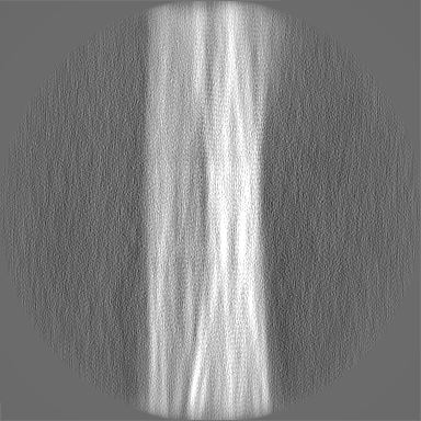
| Projections & Slices |
| ||||||||||||
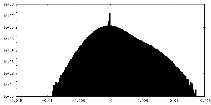
| Density Histograms |


-Half map: #1
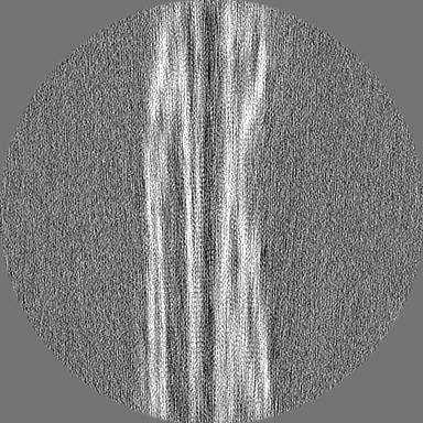
| File | emd_43826_half_map_1.map | ||||||||||||
|---|---|---|---|---|---|---|---|---|---|---|---|---|---|
| Projections & Slices |
| ||||||||||||
| Density Histograms |

-Half map: #2
| File | emd_43826_half_map_2.map | ||||||||||||
|---|---|---|---|---|---|---|---|---|---|---|---|---|---|
| Projections & Slices |
| ||||||||||||
| Density Histograms |


- Sample components
-Entire : Tau(297-407)-4E
| Entire | Name: Tau(297-407)-4E |
|---|---|
| Components |
|
-Supramolecule #1: Tau(297-407)-4E
| Supramolecule | Name: Tau(297-407)-4E / type: cell / ID: 1 / Parent: 0 / Details: tau construct with phospho-mimetic mutations |
|---|---|
| Source (natural) | Organism: |
-Experimental details
-Structure determination
| Method | cryo EM |
|---|---|
 Processing Processing | helical reconstruction |
| Aggregation state | filament |
-Sample preparation
| Buffer | pH: 7.5 |
|---|---|
| Vitrification | Cryogen name: ETHANE / Chamber humidity: 100 % / Instrument: FEI VITROBOT MARK IV |
- Electron microscopy
| Microscope | TFS KRIOS |
|---|---|
| Image recording | Film or detector model: GATAN K3 BIOQUANTUM (6k x 4k) / Average electron dose: 1.033 e/Å2 |
| Electron beam | Acceleration voltage: 300 kV / Electron source:  FIELD EMISSION GUN FIELD EMISSION GUN |
| Electron optics | Illumination mode: OTHER / Imaging mode: BRIGHT FIELD / Nominal defocus max: 2.0 µm / Nominal defocus min: 0.2 µm |
| Experimental equipment |  Model: Titan Krios / Image courtesy: FEI Company |
-Image processing
| Final reconstruction | Applied symmetry - Helical parameters - Δz: 4.8 Å Applied symmetry - Helical parameters - Δ&Phi: -0.8 ° Applied symmetry - Helical parameters - Axial symmetry: C1 (asymmetric) Resolution.type: BY AUTHOR / Resolution: 4.5 Å / Resolution method: FSC 0.143 CUT-OFF / Number images used: 85048 |
|---|---|
| CTF correction | Type: PHASE FLIPPING AND AMPLITUDE CORRECTION |
| Startup model | Type of model: NONE |
| Final angle assignment | Type: NOT APPLICABLE |
-Atomic model buiding 1
| Refinement | Protocol: AB INITIO MODEL |
|---|
