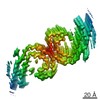
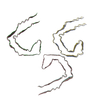
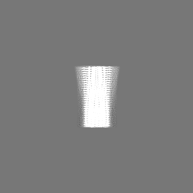
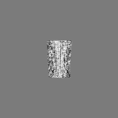
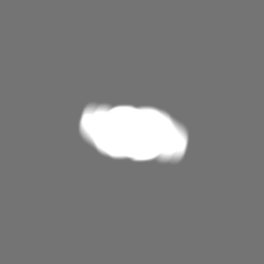
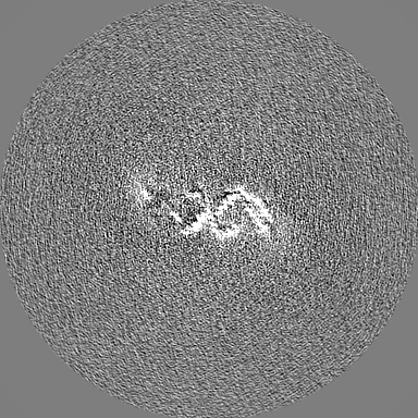
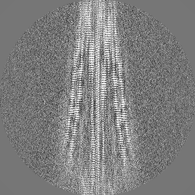
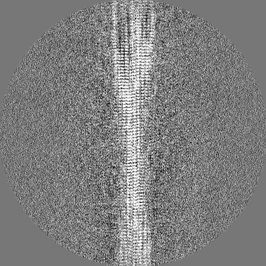
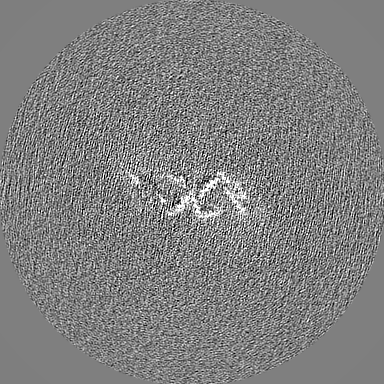
- EMDB-44422: PHF filament generated from 4E-Tau(297-407) under neutral Mg2+ co... -
+
Open data
ID or keywords:
Loading...
-
Basic information
Entry
Database: EMDB / ID: EMD-44422
Title
PHF filament generated from 4E-Tau(297-407) under neutral Mg2+ condition
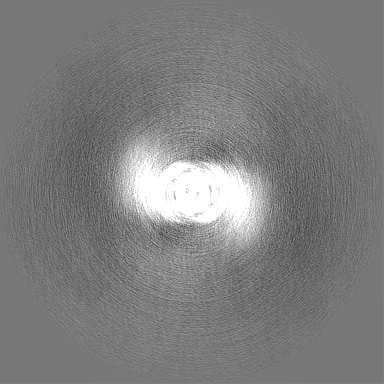
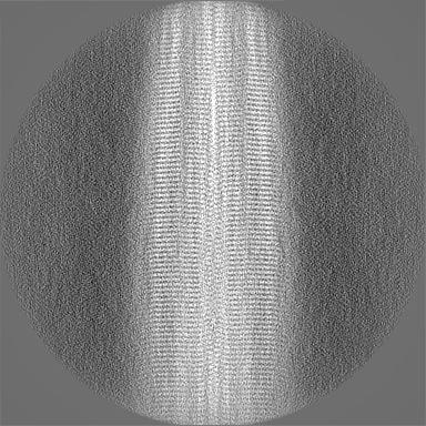
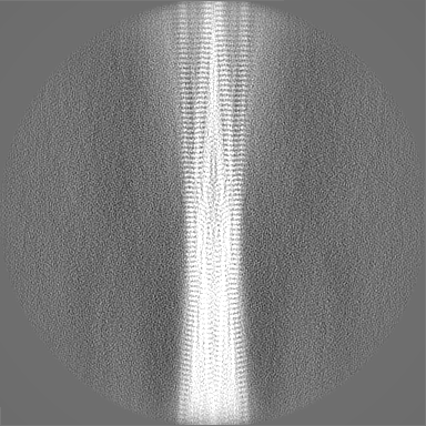
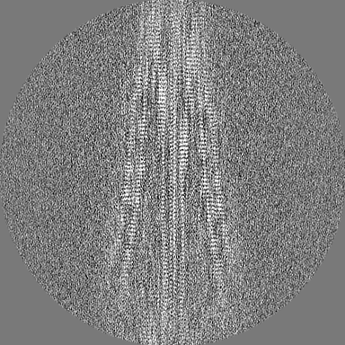
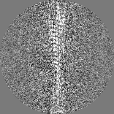
Map data
Sample
Complex: In vitro 4E-tau (297-407) PHF fibrils
Protein or peptide: Isoform Tau-F of Microtubule-associated protein tau
Keywords
Tau / amyloid fibrils / in vitro / PHF / PROTEIN FIBRIL
Function / homology
Function and homology information
plus-end-directed organelle transport along microtubule / histone-dependent DNA binding / negative regulation of establishment of protein localization to mitochondrion / neurofibrillary tangle / microtubule lateral binding / axonal transport / tubulin complex / positive regulation of protein localization to synapse / negative regulation of tubulin deacetylation / phosphatidylinositol bisphosphate binding ...plus-end-directed organelle transport along microtubule / histone-dependent DNA binding / negative regulation of establishment of protein localization to mitochondrion / neurofibrillary tangle / microtubule lateral binding / axonal transport / tubulin complex / positive regulation of protein localization to synapse / negative regulation of tubulin deacetylation / phosphatidylinositol bisphosphate binding / generation of neurons / rRNA metabolic process / axonal transport of mitochondrion / regulation of mitochondrial fission / axon development / regulation of chromosome organization / central nervous system neuron development / intracellular distribution of mitochondria / minor groove of adenine-thymine-rich DNA binding / lipoprotein particle binding / microtubule polymerization / negative regulation of mitochondrial membrane potential / dynactin binding / regulation of microtubule polymerization / apolipoprotein binding / main axon / protein polymerization / axolemma / glial cell projection / Caspase-mediated cleavage of cytoskeletal proteins / regulation of microtubule polymerization or depolymerization / negative regulation of mitochondrial fission / neurofibrillary tangle assembly / positive regulation of axon extension / regulation of cellular response to heat / Activation of AMPK downstream of NMDARs / synapse assembly / positive regulation of superoxide anion generation / regulation of long-term synaptic depression / positive regulation of protein localization / supramolecular fiber organization / cellular response to brain-derived neurotrophic factor stimulus / cytoplasmic microtubule organization / regulation of calcium-mediated signaling / positive regulation of microtubule polymerization / somatodendritic compartment / axon cytoplasm / astrocyte activation / stress granule assembly / phosphatidylinositol binding / nuclear periphery / regulation of microtubule cytoskeleton organization / protein phosphatase 2A binding / cellular response to reactive oxygen species / Hsp90 protein binding / microglial cell activation / cellular response to nerve growth factor stimulus / synapse organization / protein homooligomerization / PKR-mediated signaling / regulation of synaptic plasticity / SH3 domain binding / response to lead ion / microtubule cytoskeleton organization / memory / cytoplasmic ribonucleoprotein granule / neuron projection development / cell-cell signaling / single-stranded DNA binding / protein-folding chaperone binding / cellular response to heat / actin binding / microtubule cytoskeleton / cell body / growth cone / double-stranded DNA binding / protein-macromolecule adaptor activity / microtubule binding / dendritic spine / sequence-specific DNA binding / amyloid fibril formation / microtubule / learning or memory / neuron projection / regulation of autophagy / membrane raft / axon / negative regulation of gene expression / neuronal cell body / dendrite / DNA damage response / protein kinase binding / enzyme binding / mitochondrion / DNA binding / RNA binding / extracellular region / identical protein binding / nucleus / plasma membrane Similarity search - Function
Microtubule-associated protein Tau / Microtubule associated protein, tubulin-binding repeat / Tau and MAP protein, tubulin-binding repeat / Tau and MAP proteins tubulin-binding repeat signature. / Tau and MAP proteins tubulin-binding repeat profile. / : Similarity search - Domain/homology
National Institutes of Health/National Institute on Aging (NIH/NIA)
AG059661
United States
Citation
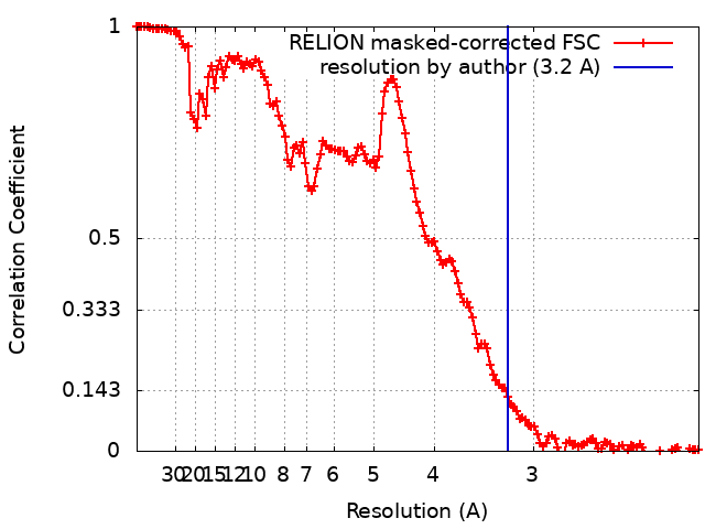
Journal: J Biol Chem / Year: 2024 Title: Milligram-scale assembly and NMR fingerprint of tau fibrils adopting the Alzheimer's disease fold. Authors: Pu Duan / Nadia El Mammeri / Mei Hong / Abstract: In the Alzheimer's disease (AD) brain, the microtubule-associated protein tau aggregates into paired helical filaments in which each protofilament has a C-shaped conformation. In vitro assembly of ...In the Alzheimer's disease (AD) brain, the microtubule-associated protein tau aggregates into paired helical filaments in which each protofilament has a C-shaped conformation. In vitro assembly of tau fibrils adopting this fold is highly valuable for both fundamental and applied studies of AD without requiring patient-brain extracted fibrils. To date, reported methods for forming AD-fold tau fibrils have been irreproducible and sensitive to subtle variations in fibrillization conditions. Here, we describe a route to reproducibly assemble tau fibrils adopting the AD fold on the multi-milligram scale. We investigated the fibrillization conditions of two constructs and found that a tau (297-407) construct that contains four AD phospho-mimetic glutamate mutations robustly formed the C-shaped conformation. 2D and 3D correlation solid-state NMR spectra show a single predominant set of chemical shifts, indicating a single molecular conformation. Negative-stain electron microscopy and cryo-EM data confirm that the protofilament formed by 4E-tau (297-407) adopts the C-shaped conformation, which associates into paired, triple, and quadruple helical filaments. In comparison, NMR spectra indicate that a previously reported construct, tau (297-391), forms a mixture of a four-layered dimer structure and the C-shaped structure, whose populations are sensitive to the environmental conditions. The determination of the NMR chemical shifts of the AD-fold tau opens the possibility for future studies of tau fibril conformations and ligand binding by NMR. The quantitative assembly of tau fibrils adopting the AD fold should facilitate the development of diagnostic and therapeutic compounds that target AD tau.
In the structure databanks used in Yorodumi, some data are registered as the other names, "COVID-19 virus" and "2019-nCoV". Here are the details of the virus and the list of structure data.
Jan 31, 2019. EMDB accession codes are about to change! (news from PDBe EMDB page)
EMDB accession codes are about to change! (news from PDBe EMDB page)
The allocation of 4 digits for EMDB accession codes will soon come to an end. Whilst these codes will remain in use, new EMDB accession codes will include an additional digit and will expand incrementally as the available range of codes is exhausted. The current 4-digit format prefixed with “EMD-” (i.e. EMD-XXXX) will advance to a 5-digit format (i.e. EMD-XXXXX), and so on. It is currently estimated that the 4-digit codes will be depleted around Spring 2019, at which point the 5-digit format will come into force.
The EM Navigator/Yorodumi systems omit the EMD- prefix.
Related info.:Q: What is EMD? / ID/Accession-code notation in Yorodumi/EM Navigator
Yorodumi is a browser for structure data from EMDB, PDB, SASBDB, etc.
This page is also the successor to EM Navigator detail page, and also detail information page/front-end page for Omokage search.
The word "yorodu" (or yorozu) is an old Japanese word meaning "ten thousand". "mi" (miru) is to see.
Related info.:EMDB / PDB / SASBDB / Comparison of 3 databanks / Yorodumi Search / Aug 31, 2016. New EM Navigator & Yorodumi / Yorodumi Papers / Jmol/JSmol / Function and homology information / Changes in new EM Navigator and Yorodumi
 Movie
Movie Controller

 Yorodumi
 Open data
 Basic information
 Map data
 Sample
 Keywords
 Function and homology information
 Homo sapiens (human)
 Authors
 United States, 1 items
  Citation
 Structure visualization
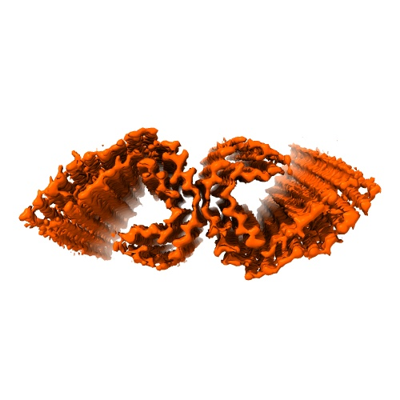
 Downloads & links
 emd_44422.png
 http://ftp.pdbj.org/pub/emdb/structures/EMD-44422


 Z (Sec.)
 Y (Row.)
 X (Col.)


 Sample components
 Processing
 Electron microscopy
 FIELD EMISSION GUN