Movie
Movie Controller


+ Open data

- Basic information
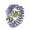
| Entry | Database: PDB / ID: 9b3f | ||||||||||||||||||
|---|---|---|---|---|---|---|---|---|---|---|---|---|---|---|---|---|---|---|---|
| Title | Cryo-EM structure of yeast (Nap1)2-H2A-H2B-Kap114 | ||||||||||||||||||
 Components Components |
| ||||||||||||||||||
 Keywords Keywords | TRANSPORT PROTEIN / Histone / Chaperone / Import / Nucleosome Assembly | ||||||||||||||||||
| Function / homology | : / : / : / :  Function and homology information Function and homology information | ||||||||||||||||||
| Biological species |  | ||||||||||||||||||
| Method | ELECTRON MICROSCOPY / single particle reconstruction / cryo EM / Resolution: 3.54 Å | ||||||||||||||||||
 Authors Authors | Jiou, J. / Fung, H.Y.J. / Chook, Y.M. | ||||||||||||||||||
| Funding support |  United States, 5items United States, 5items
| ||||||||||||||||||
 Citation Citation | Journal: J Cell Biol / Year: 2025 Title: Nap1 and Kap114 co-chaperone H2A-H2B and facilitate targeted histone release in the nucleus. Authors: Ho Yee Joyce Fung / Jenny Jiou / Ashley B Niesman / Natalia E Bernardes / Yuh Min Chook / Abstract: Core histones, synthesized and processed in the cytoplasm, must be chaperoned as they are transported into the nucleus for nucleosome assembly. The importin Kap114 transports H2A-H2B into the yeast ...Core histones, synthesized and processed in the cytoplasm, must be chaperoned as they are transported into the nucleus for nucleosome assembly. The importin Kap114 transports H2A-H2B into the yeast nucleus, where RanGTP facilitates histone release. Kap114 and H2A-H2B also bind the histone chaperone Nap1, but how Nap1 and Kap114 cooperate in transport and nucleosome assembly remains unclear. Here, biochemical and structural analyses show that Kap114, H2A-H2B, and a Nap1 dimer (Nap12) associate in the absence and presence of RanGTP to form equimolar complexes. A previous study had shown that RanGTP reduces Kap114's ability to chaperone H2A-H2B, but a new cryo-EM structure of the Nap12•H2A-H2B•Kap114•RanGTP complex explains how both Kap114 and Nap12 interact with H2A-H2B, restoring its chaperoning within the assembly while effectively depositing it into nucleosomes. Together, our results suggest that Kap114 and Nap12 provide a sheltered path that facilitates the transfer of H2A-H2B from Kap114 to Nap12, ultimately directing its specific deposition into nucleosomes. | ||||||||||||||||||
| History |
|
- Structure visualization
| Structure viewer | Molecule: MolmilJmol/JSmol |
|---|
- Downloads & links
-Download
| PDBx/mmCIF format | 9b3f.cif.gz | 563.3 KB | Display | PDBx/mmCIF format |
|---|---|---|---|---|
| PDB format | pdb9b3f.ent.gz | 463.1 KB | Display | PDB format |
| PDBx/mmJSON format | 9b3f.json.gz | Tree view | PDBx/mmJSON format | |
| Others |  Other downloads Other downloads |
-Validation report
| Arichive directory | https://data.pdbj.org/pub/pdb/validation_reports/b3/9b3fftp://data.pdbj.org/pub/pdb/validation_reports/b3/9b3f | HTTPS FTP |
|---|
-Related structure data
| Related structure data |  44136MC  9b23C  9b31C  9b3iC C: citing same article ( M: map data used to model this data |
|---|---|
| Similar structure data |


-Links
 PDBj
PDBj

- Assembly
| Deposited unit | 
|
|---|---|
| 1 |
|
-Components
| #1: Protein | Mass: 36182.355 Da / Num. of mol.: 2 Source method: isolated from a genetically manipulated source Source: (gene. exp.) Gene: NAP1, GI527_G0003692 / Production host:  #2: Protein | | Mass: 114220.867 Da / Num. of mol.: 1 Source method: isolated from a genetically manipulated source Source: (gene. exp.) Gene: KAP114, GI527_G0002177 / Production host: #3: Protein | | Mass: 13881.980 Da / Num. of mol.: 1 Source method: isolated from a genetically manipulated source Source: (gene. exp.) Gene: HTA2, GI527_G0000202 / Production host: #4: Protein | | Mass: 14133.145 Da / Num. of mol.: 1 Source method: isolated from a genetically manipulated source Source: (gene. exp.) Gene: HTB2, GI527_G0000203 / Production host: Has protein modification | N | |
|---|
-Experimental details
-Experiment
| Experiment | Method: ELECTRON MICROSCOPY |
|---|---|
| EM experiment | Aggregation state: PARTICLE / 3D reconstruction method: single particle reconstruction |
- Sample preparation
| Component | Name: Crosslinked mixture of Kap114 with Nap1 and H2A-H2B / Type: COMPLEX Details: Nap1 and H2A-H2B were pre-complexed with excess histones, and Kap114 was added before mild crosslinking and SEC separation. Entity ID: all / Source: RECOMBINANT | |||||||||||||||||||||||||
|---|---|---|---|---|---|---|---|---|---|---|---|---|---|---|---|---|---|---|---|---|---|---|---|---|---|---|
| Molecular weight | Value: 0.072 MDa / Experimental value: NO | |||||||||||||||||||||||||
| Source (natural) | Organism: | |||||||||||||||||||||||||
| Source (recombinant) | Organism: | |||||||||||||||||||||||||
| Buffer solution | pH: 7.4 | |||||||||||||||||||||||||
| Buffer component |
| |||||||||||||||||||||||||
| Specimen | Conc.: 1.2 mg/ml / Embedding applied: NO / Shadowing applied: NO / Staining applied: NO / Vitrification applied: YES / Details: Crosslinked sample. | |||||||||||||||||||||||||
| Specimen support | Grid material: COPPER / Grid type: Quantifoil | |||||||||||||||||||||||||
| Vitrification | Instrument: FEI VITROBOT MARK IV / Cryogen name: ETHANE / Humidity: 95 % / Chamber temperature: 280 K |
- Electron microscopy imaging
| Experimental equipment |  Model: Titan Krios / Image courtesy: FEI Company |
|---|---|
| Microscopy | Model: FEI TITAN KRIOS |
| Electron gun | Electron source:  FIELD EMISSION GUN / Accelerating voltage: 300 kV / Illumination mode: FLOOD BEAM FIELD EMISSION GUN / Accelerating voltage: 300 kV / Illumination mode: FLOOD BEAM |
| Electron lens | Mode: BRIGHT FIELD / Nominal magnification: 105000 X / Nominal defocus max: 2400 nm / Nominal defocus min: 900 nm |
| Specimen holder | Specimen holder model: FEI TITAN KRIOS AUTOGRID HOLDER |
| Image recording | Average exposure time: 5.4 sec. / Electron dose: 59 e/Å2 / Film or detector model: GATAN K3 (6k x 4k) / Num. of grids imaged: 1 / Num. of real images: 1080 |
| EM imaging optics | Energyfilter slit width: 20 eV |
- Processing
| EM software |
| |||||||||||||||||||||||||||||||||||||||||||||||||||||||
|---|---|---|---|---|---|---|---|---|---|---|---|---|---|---|---|---|---|---|---|---|---|---|---|---|---|---|---|---|---|---|---|---|---|---|---|---|---|---|---|---|---|---|---|---|---|---|---|---|---|---|---|---|---|---|---|---|
| CTF correction | Type: PHASE FLIPPING AND AMPLITUDE CORRECTION | |||||||||||||||||||||||||||||||||||||||||||||||||||||||
| Particle selection | Num. of particles selected: 4314112 Details: Blob picking on 100 micrographs and then further template picking. | |||||||||||||||||||||||||||||||||||||||||||||||||||||||
| 3D reconstruction | Resolution: 3.54 Å / Resolution method: FSC 0.143 CUT-OFF / Num. of particles: 113011 Details: Composite map was generated by first multiplying the consensus volume with the particle subtraction mask using vop multiply, and composited with the locally refined map covering the Nap1 and ...Details: Composite map was generated by first multiplying the consensus volume with the particle subtraction mask using vop multiply, and composited with the locally refined map covering the Nap1 and histone area by vop maximum in ChimeraX. Symmetry type: POINT | |||||||||||||||||||||||||||||||||||||||||||||||||||||||
| Atomic model building | Protocol: OTHER | |||||||||||||||||||||||||||||||||||||||||||||||||||||||
| Atomic model building | 3D fitting-ID: 1
| |||||||||||||||||||||||||||||||||||||||||||||||||||||||
| Refine LS restraints |
|