National Institutes of Health/National Institute of General Medical Sciences (NIH/NIGMS)
R35GM141461
United States
National Institutes of Health/National Institute of General Medical Sciences (NIH/NIGMS)
R01GM069909
United States
National Institutes of Health/National Institute of General Medical Sciences (NIH/NIGMS)
T32GM008203
United States
Welch Foundation
I-1532
United States
Cancer Prevention and Research Institute of Texas (CPRIT)
RP220582
United States
Citation
Journal: J Cell Biol / Year: 2025 Title: Nap1 and Kap114 co-chaperone H2A-H2B and facilitate targeted histone release in the nucleus. Authors: Ho Yee Joyce Fung / Jenny Jiou / Ashley B Niesman / Natalia E Bernardes / Yuh Min Chook / Abstract: Core histones, synthesized and processed in the cytoplasm, must be chaperoned as they are transported into the nucleus for nucleosome assembly. The importin Kap114 transports H2A-H2B into the yeast ...Core histones, synthesized and processed in the cytoplasm, must be chaperoned as they are transported into the nucleus for nucleosome assembly. The importin Kap114 transports H2A-H2B into the yeast nucleus, where RanGTP facilitates histone release. Kap114 and H2A-H2B also bind the histone chaperone Nap1, but how Nap1 and Kap114 cooperate in transport and nucleosome assembly remains unclear. Here, biochemical and structural analyses show that Kap114, H2A-H2B, and a Nap1 dimer (Nap12) associate in the absence and presence of RanGTP to form equimolar complexes. A previous study had shown that RanGTP reduces Kap114's ability to chaperone H2A-H2B, but a new cryo-EM structure of the Nap12•H2A-H2B•Kap114•RanGTP complex explains how both Kap114 and Nap12 interact with H2A-H2B, restoring its chaperoning within the assembly while effectively depositing it into nucleosomes. Together, our results suggest that Kap114 and Nap12 provide a sheltered path that facilitates the transfer of H2A-H2B from Kap114 to Nap12, ultimately directing its specific deposition into nucleosomes.
Entire : Complex of Kap114 bound to Ran GTPase Gsp1, H2A-H2B and Nap1
Entire
Name: Complex of Kap114 bound to Ran GTPase Gsp1, H2A-H2B and Nap1
Components
Complex: Complex of Kap114 bound to Ran GTPase Gsp1, H2A-H2B and Nap1
-
Supramolecule #1: Complex of Kap114 bound to Ran GTPase Gsp1, H2A-H2B and Nap1
Supramolecule
Name: Complex of Kap114 bound to Ran GTPase Gsp1, H2A-H2B and Nap1 type: complex / ID: 1 / Parent: 0 / Macromolecule list: #1-#6 Details: Complex was formed and dialyzed overnight, then mildly crosslinked and separated by size-exclusion chromatography.
Model: Quantifoil R2/1 / Material: COPPER / Mesh: 300 / Support film - Material: CARBON / Support film - topology: HOLEY / Pretreatment - Type: GLOW DISCHARGE / Pretreatment - Time: 80 sec.
Vitrification
Cryogen name: ETHANE / Chamber humidity: 95 % / Chamber temperature: 280 K / Instrument: FEI VITROBOT MARK IV
Details
Crosslinked sample.
-
Electron microscopy
Microscope
FEI TITAN KRIOS
Specialist optics
Energy filter - Slit width: 20 eV
Image recording
Film or detector model: FEI FALCON IV (4k x 4k) / Number grids imaged: 1 / Number real images: 9331 / Average exposure time: 3.6 sec. / Average electron dose: 50.0 e/Å2
Electron beam
Acceleration voltage: 300 kV / Electron source: FIELD EMISSION GUN
Number selected: 1381753 / Details: Blob picker then followed by Topaz picking.
Startup model
Type of model: NONE / Details: Ab-initio reconstruction.
Final reconstruction
Resolution.type: BY AUTHOR / Resolution: 3.97 Å / Resolution method: FSC 0.143 CUT-OFF / Software - Name: cryoSPARC Details: Particle subtraction was performed with a mask covering RanGTP and Kap114, and local refinement was performed for Nap1 and H2A-H2B bound region. Number images used: 133516
Initial angle assignment
Type: MAXIMUM LIKELIHOOD / Software - Name: cryoSPARC
Final angle assignment
Type: MAXIMUM LIKELIHOOD / Software - Name: cryoSPARC
Final 3D classification
Software - Name: cryoSPARC
FSC plot (resolution estimation)
+
About Yorodumi
-
News
-
Feb 9, 2022. New format data for meta-information of EMDB entries
New format data for meta-information of EMDB entries
Version 3 of the EMDB header file is now the official format.
The previous official version 1.9 will be removed from the archive.
In the structure databanks used in Yorodumi, some data are registered as the other names, "COVID-19 virus" and "2019-nCoV". Here are the details of the virus and the list of structure data.
Jan 31, 2019. EMDB accession codes are about to change! (news from PDBe EMDB page)
EMDB accession codes are about to change! (news from PDBe EMDB page)
The allocation of 4 digits for EMDB accession codes will soon come to an end. Whilst these codes will remain in use, new EMDB accession codes will include an additional digit and will expand incrementally as the available range of codes is exhausted. The current 4-digit format prefixed with “EMD-” (i.e. EMD-XXXX) will advance to a 5-digit format (i.e. EMD-XXXXX), and so on. It is currently estimated that the 4-digit codes will be depleted around Spring 2019, at which point the 5-digit format will come into force.
The EM Navigator/Yorodumi systems omit the EMD- prefix.
Related info.:Q: What is EMD? / ID/Accession-code notation in Yorodumi/EM Navigator
Yorodumi is a browser for structure data from EMDB, PDB, SASBDB, etc.
This page is also the successor to EM Navigator detail page, and also detail information page/front-end page for Omokage search.
The word "yorodu" (or yorozu) is an old Japanese word meaning "ten thousand". "mi" (miru) is to see.
Related info.:EMDB / PDB / SASBDB / Comparison of 3 databanks / Yorodumi Search / Aug 31, 2016. New EM Navigator & Yorodumi / Yorodumi Papers / Jmol/JSmol / Function and homology information / Changes in new EM Navigator and Yorodumi
 Movie
Movie Controller

 Yorodumi
 Open data
 Basic information
 Map data
 Sample
 Keywords
 Authors
 United States, 5 items
  Citation
 Structure visualization
 Downloads & links
 EMDB map data format
 emd_44150.png
 http://ftp.pdbj.org/pub/emdb/structures/EMD-44150
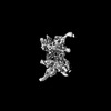


 Z (Sec.)
 Y (Row.)
 X (Col.)


 Sample components
 Processing
 Electron microscopy
 FIELD EMISSION GUN

