 Movie
Movie Controller

[English] 日本語
 Yorodumi
- PDB-8znc: Cryo-EM structure of W89F mutated Glutamate dehydrogenase from Th... -
+ Open data

- Basic information
| Entry | Database: PDB / ID: 8znc | ||||||||||||||||||||||||||||||||||||||||||
|---|---|---|---|---|---|---|---|---|---|---|---|---|---|---|---|---|---|---|---|---|---|---|---|---|---|---|---|---|---|---|---|---|---|---|---|---|---|---|---|---|---|---|---|
| Title | Cryo-EM structure of W89F mutated Glutamate dehydrogenase from Thermococcus profundus incorporating NADPH, AKG in the steady stage of reaction | ||||||||||||||||||||||||||||||||||||||||||
 Components Components | Glutamate dehydrogenase | ||||||||||||||||||||||||||||||||||||||||||
 Keywords Keywords | OXIDOREDUCTASE / Complex / NADPH / 2-oxoglutarate / Mutant | ||||||||||||||||||||||||||||||||||||||||||
| Function / homology |  Function and homology information Function and homology informationglutamate dehydrogenase [NAD(P)+] / L-glutamate dehydrogenase (NADP+) activity / L-glutamate dehydrogenase (NAD+) activity / L-glutamate catabolic process Similarity search - Function | ||||||||||||||||||||||||||||||||||||||||||
| Biological species |   Thermococcus profundus (archaea) Thermococcus profundus (archaea) | ||||||||||||||||||||||||||||||||||||||||||
| Method | ELECTRON MICROSCOPY / single particle reconstruction / cryo EM / Resolution: 2.41 Å | ||||||||||||||||||||||||||||||||||||||||||
 Authors Authors | Wakabayashi, T. / Nakasako, M. | ||||||||||||||||||||||||||||||||||||||||||
| Funding support |  Japan, 13items Japan, 13items
| ||||||||||||||||||||||||||||||||||||||||||
 Citation Citation | Journal: FEBS J / Year: 2025 Title: CryoEM and crystal structure analyses reveal the indirect role played by Trp89 in glutamate dehydrogenase enzymatic reactions. Authors: Taiki Wakabayashi / Yuka Matsui / Masayoshi Nakasako / Abstract: Glutamate dehydrogenase from Thermococcus profundus is a homo-hexameric enzyme that catalyzes the reversible deamination of glutamate to 2-oxoglutarate in the presence of a cofactor. In each subunit, ...Glutamate dehydrogenase from Thermococcus profundus is a homo-hexameric enzyme that catalyzes the reversible deamination of glutamate to 2-oxoglutarate in the presence of a cofactor. In each subunit, a large active-site cleft is formed between the two functional domains, one of which displays motion to open and close the cleft. Trp89 in the cleft displays two sidechain conformers in the open cleft and a single conformer in the closed cleft. To reveal the role of the Trp89 sidechain in the domain motion, we mutated Trp89 to phenylalanine. Despite the Trp89 sidechain being located away from the reaction center, the catalytic constant decreased to 1/38-fold of that of the wild-type without a fatal reduction of the affinities to the cofactor and ligand molecules. To understand the molecular mechanism underlying this reduction, we determined the crystal structure in the unliganded state and the metastable conformations appearing in the steady stage of the reaction using cryo-electron microscopy (cryoEM). The four identified metastable conformations were similar to the three conformations observed in the wild-type, but their populations were different from those of the wild-type. In addition, a conformation with a completely closed active-site cleft necessary for the reaction to proceed was quite rare. The crystal structure and the four metastable conformations suggested that the reduction in the catalytic constant could be attributed to changes in the interactions between Gln13 and the 89th side chains, preventing the closing domain motion. | ||||||||||||||||||||||||||||||||||||||||||
| History |
|
- Structure visualization
| Structure viewer | Molecule: MolmilJmol/JSmol |
|---|
- Downloads & links
-Download
| PDBx/mmCIF format | 8znc.cif.gz | 86.3 KB | Display | PDBx/mmCIF format |
|---|---|---|---|---|
| PDB format | pdb8znc.ent.gz | 62.3 KB | Display | PDB format |
| PDBx/mmJSON format | 8znc.json.gz | Tree view | PDBx/mmJSON format | |
| Others |  Other downloads Other downloads |
-Validation report
| Arichive directory | https://data.pdbj.org/pub/pdb/validation_reports/zn/8zncftp://data.pdbj.org/pub/pdb/validation_reports/zn/8znc | HTTPS FTP |
|---|
-Related structure data
| Related structure data |  60267MC  8zmuC  8znbC 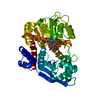 8zndC 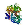 8zneC  8zngC C: citing same article ( M: map data used to model this data |
|---|---|
| Similar structure data |


-Links
 PDBj
PDBj


- Assembly
| Deposited unit | 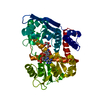
|
|---|---|
| 1 |
|
-Components
| #1: Protein | Mass: 46360.004 Da / Num. of mol.: 1 / Mutation: W89F Source method: isolated from a genetically manipulated source Source: (gene. exp.) Thermococcus profundus (archaea) / Gene: gdhA, gdh / Production host:  References: UniProt: O74024, glutamate dehydrogenase [NAD(P)+] |
|---|---|
| #2: Chemical | ChemComp-NDP /   Mass: 745.421 Da / Num. of mol.: 1 / Source method: obtained synthetically / Formula: C21H30N7O17P3 Mass: 745.421 Da / Num. of mol.: 1 / Source method: obtained synthetically / Formula: C21H30N7O17P3 |
| #3: Chemical | ChemComp-AKG /   Mass: 146.098 Da / Num. of mol.: 1 / Source method: obtained synthetically / Formula: C5H6O5 / Feature type: SUBJECT OF INVESTIGATION Mass: 146.098 Da / Num. of mol.: 1 / Source method: obtained synthetically / Formula: C5H6O5 / Feature type: SUBJECT OF INVESTIGATION |
| #4: Chemical | ChemComp-NH4 /   Mass: 18.038 Da / Num. of mol.: 1 / Source method: obtained synthetically / Formula: H4N / Feature type: SUBJECT OF INVESTIGATION Mass: 18.038 Da / Num. of mol.: 1 / Source method: obtained synthetically / Formula: H4N / Feature type: SUBJECT OF INVESTIGATION |
| #5: Water | ChemComp-HOH /  Mass: 18.015 Da / Num. of mol.: 24 / Source method: isolated from a natural source / Formula: H2O Mass: 18.015 Da / Num. of mol.: 24 / Source method: isolated from a natural source / Formula: H2O |
| Has ligand of interest | Y |
| Has protein modification | N |
-Experimental details
-Experiment
| Experiment | Method: ELECTRON MICROSCOPY |
|---|---|
| EM experiment | Aggregation state: PARTICLE / 3D reconstruction method: single particle reconstruction |
- Sample preparation
| Component | Name: Hexamer of W89F mutated glutamate dehydrogenase / Type: ORGANELLE OR CELLULAR COMPONENT / Entity ID: #1 / Source: RECOMBINANT | ||||||||||||||||||||
|---|---|---|---|---|---|---|---|---|---|---|---|---|---|---|---|---|---|---|---|---|---|
| Molecular weight | Value: 0.280 MDa / Experimental value: YES | ||||||||||||||||||||
| Source (natural) | Organism: Thermococcus profundus (archaea) | ||||||||||||||||||||
| Source (recombinant) | Organism: | ||||||||||||||||||||
| Buffer solution | pH: 7.5 Details: 0.5 mM NADP, 100 mM Glutamate in 5 mM Tris-HCl at pH7.5. | ||||||||||||||||||||
| Buffer component |
| ||||||||||||||||||||
| Specimen | Conc.: 9 mg/ml / Embedding applied: NO / Shadowing applied: NO / Staining applied: NO / Vitrification applied: YES / Details: 8.96 mg/mL GDH W89F | ||||||||||||||||||||
| Specimen support | Details: Both sides of the grid were glow-discharged for 45 s at 20 mA and 20 Pa. Grid material: COPPER / Grid mesh size: 200 divisions/in. / Grid type: Quantifoil R1.2/1.3 | ||||||||||||||||||||
| Vitrification | Instrument: FEI VITROBOT MARK IV / Cryogen name: ETHANE / Humidity: 100 % / Chamber temperature: 281 K Details: The sample solution was flash-frozen 2-h after mixing the protein solution and buffer solution. |
- Electron microscopy imaging
| Microscopy | Model: JEOL CRYO ARM 300 |
|---|---|
| Electron gun | Electron source:  FIELD EMISSION GUN / Accelerating voltage: 300 kV / Illumination mode: OTHER FIELD EMISSION GUN / Accelerating voltage: 300 kV / Illumination mode: OTHER |
| Electron lens | Mode: BRIGHT FIELD / Nominal defocus max: 5000 nm / Nominal defocus min: 350 nm |
| Specimen holder | Cryogen: NITROGEN |
| Image recording | Average exposure time: 2 sec. / Electron dose: 50 e/Å2 / Film or detector model: GATAN K3 (6k x 4k) / Num. of grids imaged: 1 / Num. of real images: 7651 |
- Processing
| EM software |
| ||||||||||||||||||||||||||||||||||||||||
|---|---|---|---|---|---|---|---|---|---|---|---|---|---|---|---|---|---|---|---|---|---|---|---|---|---|---|---|---|---|---|---|---|---|---|---|---|---|---|---|---|---|
| CTF correction | Type: PHASE FLIPPING AND AMPLITUDE CORRECTION | ||||||||||||||||||||||||||||||||||||||||
| Particle selection | Num. of particles selected: 3534400 | ||||||||||||||||||||||||||||||||||||||||
| 3D reconstruction | Resolution: 2.41 Å / Resolution method: FSC 0.143 CUT-OFF / Num. of particles: 444903 / Num. of class averages: 1 / Symmetry type: POINT |
