 Movie
Movie Controller


+ Open data

- Basic information
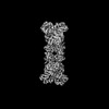
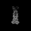
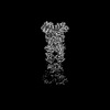
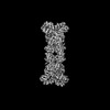
| Entry | Database: PDB / ID: 8zc9 | ||||||||||||
|---|---|---|---|---|---|---|---|---|---|---|---|---|---|
| Title | The Cryo-EM structure of DSR2-Tail tube-NAD+ complex | ||||||||||||
 Components Components |
| ||||||||||||
 Keywords Keywords | IMMUNE SYSTEM / NADase / anti-phage defense / tail-tube protein / NAD | ||||||||||||
| Function / homology | SIR2-like domain / SIR2-like domain / DHS-like NAD/FAD-binding domain superfamily / NICOTINAMIDE-ADENINE-DINUCLEOTIDE / Uncharacterized protein / SIR2-like domain-containing protein Function and homology information Function and homology information | ||||||||||||
| Biological species |  | ||||||||||||
| Method | ELECTRON MICROSCOPY / single particle reconstruction / cryo EM / Resolution: 3.14 Å | ||||||||||||
 Authors Authors | Wang, R. / Xu, Q. / Wu, Z. / Li, J. / Yang, R. / Shi, Z. / Li, F. | ||||||||||||
| Funding support |  China, 3items China, 3items
| ||||||||||||
 Citation Citation | Journal: Nat Commun / Year: 2024 Title: The structural basis of the activation and inhibition of DSR2 NADase by phage proteins. Authors: Ruiwen Wang / Qi Xu / Zhuoxi Wu / Jialu Li / Hao Guo / Tianzhui Liao / Yuan Shi / Ling Yuan / Haishan Gao / Rong Yang / Zhubing Shi / Faxiang Li / Abstract: DSR2, a Sir2 domain-containing protein, protects bacteria from phage infection by hydrolyzing NAD. The enzymatic activity of DSR2 is triggered by the SPR phage tail tube protein (TTP), while ...DSR2, a Sir2 domain-containing protein, protects bacteria from phage infection by hydrolyzing NAD. The enzymatic activity of DSR2 is triggered by the SPR phage tail tube protein (TTP), while suppressed by the SPbeta phage-encoded DSAD1 protein, enabling phages to evade the host defense. However, the molecular mechanisms of activation and inhibition of DSR2 remain elusive. Here, we report the cryo-EM structures of apo DSR2, DSR2-TTP-NAD and DSR2-DSAD1 complexes. DSR2 assembles into a head-to-head tetramer mediated by its Sir2 domain. The C-terminal helical regions of DSR2 constitute four partner-binding cavities with opened and closed conformation. Two TTP molecules bind to two of the four C-terminal cavities, inducing conformational change of Sir2 domain to activate DSR2. Furthermore, DSAD1 competes with the activator for binding to the C-terminal cavity of DSR2, effectively suppressing its enzymatic activity. Our results provide the mechanistic insights into the DSR2-mediated anti-phage defense system and DSAD1-dependent phage immune evasion. | ||||||||||||
| History |
|
- Structure visualization
| Structure viewer | Molecule: MolmilJmol/JSmol |
|---|
- Downloads & links
-Download
| PDBx/mmCIF format | 8zc9.cif.gz | 1 MB | Display | PDBx/mmCIF format |
|---|---|---|---|---|
| PDB format | pdb8zc9.ent.gz | 706.6 KB | Display | PDB format |
| PDBx/mmJSON format | 8zc9.json.gz | Tree view | PDBx/mmJSON format | |
| Others |  Other downloads Other downloads |
-Validation report
| Arichive directory | https://data.pdbj.org/pub/pdb/validation_reports/zc/8zc9ftp://data.pdbj.org/pub/pdb/validation_reports/zc/8zc9 | HTTPS FTP |
|---|
-Related structure data
| Related structure data | 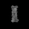 39925MC 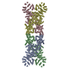 8y13C 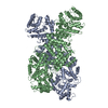 8y34C 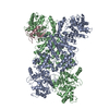 8y3mC  8y3wC  8y3yC C: citing same article ( M: map data used to model this data |
|---|---|
| Similar structure data |


-Links
 PDBj
PDBj

- Assembly
| Deposited unit | 
|
|---|---|
| 1 |
|
-Components
| #1: Protein | Mass: 118568.727 Da / Num. of mol.: 4 Source method: isolated from a genetically manipulated source Details: The sequence of organism Bacillus subtilis is not available, replaced by D4G637 temporarily. Source: (gene. exp.) #2: Protein | Mass: 29304.701 Da / Num. of mol.: 2 Source method: isolated from a genetically manipulated source Source: (gene. exp.) #3: Chemical | ChemComp-NAD /   Mass: 663.425 Da / Num. of mol.: 4 / Source method: obtained synthetically / Formula: C21H27N7O14P2 / Feature type: SUBJECT OF INVESTIGATION / Comment: NAD*YM Mass: 663.425 Da / Num. of mol.: 4 / Source method: obtained synthetically / Formula: C21H27N7O14P2 / Feature type: SUBJECT OF INVESTIGATION / Comment: NAD*YMHas ligand of interest | Y | |
|---|
-Experimental details
-Experiment
| Experiment | Method: ELECTRON MICROSCOPY |
|---|---|
| EM experiment | Aggregation state: PARTICLE / 3D reconstruction method: single particle reconstruction |
- Sample preparation
| Component | Name: DSR2-tail tube protein-NAD+ complex / Type: COMPLEX / Entity ID: #1-#2 / Source: RECOMBINANT | ||||||||||||||||||||
|---|---|---|---|---|---|---|---|---|---|---|---|---|---|---|---|---|---|---|---|---|---|
| Source (natural) | Organism: | ||||||||||||||||||||
| Source (recombinant) | Organism: | ||||||||||||||||||||
| Buffer solution | pH: 7.5 / Details: 20 mM HEPES pH 7.5, 100 mM NaCl, 0.5 mM TCEP | ||||||||||||||||||||
| Buffer component |
| ||||||||||||||||||||
| Specimen | Embedding applied: NO / Shadowing applied: NO / Staining applied: NO / Vitrification applied: YES / Details: 10mg/ml | ||||||||||||||||||||
| Specimen support | Grid material: COPPER / Grid mesh size: 300 divisions/in. / Grid type: Quantifoil R1.2/1.3 | ||||||||||||||||||||
| Vitrification | Instrument: FEI VITROBOT MARK I / Cryogen name: ETHANE / Humidity: 100 % |
- Electron microscopy imaging
| Experimental equipment |  Model: Titan Krios / Image courtesy: FEI Company |
|---|---|
| Microscopy | Model: FEI TITAN KRIOS |
| Electron gun | Electron source:  FIELD EMISSION GUN / Accelerating voltage: 300 kV / Illumination mode: FLOOD BEAM FIELD EMISSION GUN / Accelerating voltage: 300 kV / Illumination mode: FLOOD BEAM |
| Electron lens | Mode: BRIGHT FIELD / Nominal magnification: 105000 X / Nominal defocus max: 2000 nm / Nominal defocus min: 1500 nm / Cs: 2.7 mm / C2 aperture diameter: 50 µm |
| Specimen holder | Specimen holder model: FEI TITAN KRIOS AUTOGRID HOLDER |
| Image recording | Electron dose: 50 e/Å2 / Film or detector model: FEI FALCON IV (4k x 4k) |
- Processing
| CTF correction | Type: NONE | ||||||||||||||||||||||||
|---|---|---|---|---|---|---|---|---|---|---|---|---|---|---|---|---|---|---|---|---|---|---|---|---|---|
| Particle selection | Num. of particles selected: 3383 | ||||||||||||||||||||||||
| 3D reconstruction | Resolution: 3.14 Å / Resolution method: FSC 0.143 CUT-OFF / Num. of particles: 57864 / Symmetry type: POINT | ||||||||||||||||||||||||
| Atomic model building | B value: 116 | ||||||||||||||||||||||||
| Refinement | Cross valid method: NONE Stereochemistry target values: GeoStd + Monomer Library + CDL v1.2 | ||||||||||||||||||||||||
| Displacement parameters | Biso mean: 126.31 Å2 | ||||||||||||||||||||||||
| Refine LS restraints |
|
