National Natural Science Foundation of China (NSFC)
32370742
China
National Natural Science Foundation of China (NSFC)
32271264
China
National Natural Science Foundation of China (NSFC)
32200129
China
Citation
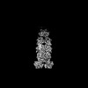
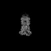
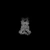
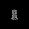
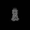
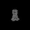
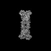
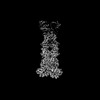
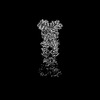
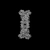
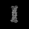
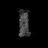
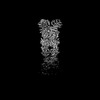
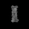
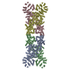
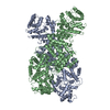
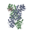
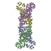
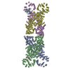
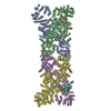
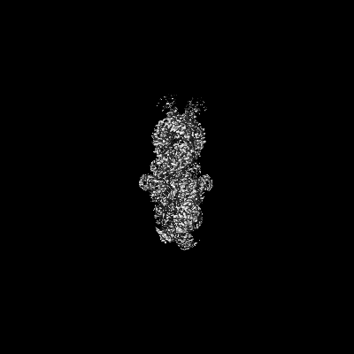
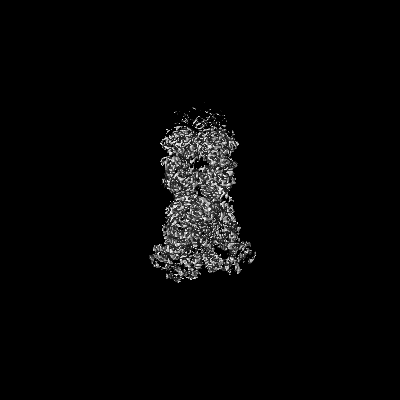
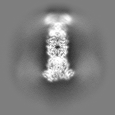
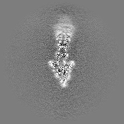
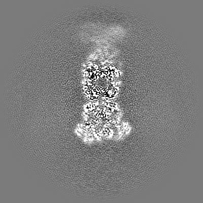
Journal: Nat Commun / Year: 2024 Title: The structural basis of the activation and inhibition of DSR2 NADase by phage proteins. Authors: Ruiwen Wang / Qi Xu / Zhuoxi Wu / Jialu Li / Hao Guo / Tianzhui Liao / Yuan Shi / Ling Yuan / Haishan Gao / Rong Yang / Zhubing Shi / Faxiang Li / Abstract: DSR2, a Sir2 domain-containing protein, protects bacteria from phage infection by hydrolyzing NAD. The enzymatic activity of DSR2 is triggered by the SPR phage tail tube protein (TTP), while ...DSR2, a Sir2 domain-containing protein, protects bacteria from phage infection by hydrolyzing NAD. The enzymatic activity of DSR2 is triggered by the SPR phage tail tube protein (TTP), while suppressed by the SPbeta phage-encoded DSAD1 protein, enabling phages to evade the host defense. However, the molecular mechanisms of activation and inhibition of DSR2 remain elusive. Here, we report the cryo-EM structures of apo DSR2, DSR2-TTP-NAD and DSR2-DSAD1 complexes. DSR2 assembles into a head-to-head tetramer mediated by its Sir2 domain. The C-terminal helical regions of DSR2 constitute four partner-binding cavities with opened and closed conformation. Two TTP molecules bind to two of the four C-terminal cavities, inducing conformational change of Sir2 domain to activate DSR2. Furthermore, DSAD1 competes with the activator for binding to the C-terminal cavity of DSR2, effectively suppressing its enzymatic activity. Our results provide the mechanistic insights into the DSR2-mediated anti-phage defense system and DSAD1-dependent phage immune evasion.
In the structure databanks used in Yorodumi, some data are registered as the other names, "COVID-19 virus" and "2019-nCoV". Here are the details of the virus and the list of structure data.
Jan 31, 2019. EMDB accession codes are about to change! (news from PDBe EMDB page)
EMDB accession codes are about to change! (news from PDBe EMDB page)
The allocation of 4 digits for EMDB accession codes will soon come to an end. Whilst these codes will remain in use, new EMDB accession codes will include an additional digit and will expand incrementally as the available range of codes is exhausted. The current 4-digit format prefixed with “EMD-” (i.e. EMD-XXXX) will advance to a 5-digit format (i.e. EMD-XXXXX), and so on. It is currently estimated that the 4-digit codes will be depleted around Spring 2019, at which point the 5-digit format will come into force.
The EM Navigator/Yorodumi systems omit the EMD- prefix.
Related info.:Q: What is EMD? / ID/Accession-code notation in Yorodumi/EM Navigator
Yorodumi is a browser for structure data from EMDB, PDB, SASBDB, etc.
This page is also the successor to EM Navigator detail page, and also detail information page/front-end page for Omokage search.
The word "yorodu" (or yorozu) is an old Japanese word meaning "ten thousand". "mi" (miru) is to see.
Related info.:EMDB / PDB / SASBDB / Comparison of 3 databanks / Yorodumi Search / Aug 31, 2016. New EM Navigator & Yorodumi / Yorodumi Papers / Jmol/JSmol / Function and homology information / Changes in new EM Navigator and Yorodumi
 Movie
Movie Controller


 Open data
 Basic information
 Map data
 Sample
 Keywords
 Authors
 China, 3 items
  Citation
 Structure visualization
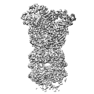
 Downloads & links
 EMDB map data format
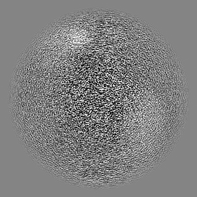
 emd_38888.png
 http://ftp.pdbj.org/pub/emdb/structures/EMD-38888


 Z (Sec.)
 Y (Row.)
 X (Col.)


 Sample components
 Processing
 Electron microscopy
 FIELD EMISSION GUN
