 Movie
Movie Controller


+ Open data

- Basic information
| Entry | Database: PDB / ID: 8z9k | ||||||
|---|---|---|---|---|---|---|---|
| Title | Crystal structure of LINE-1 ribozyme | ||||||
 Components Components | RNA (41-MER) | ||||||
 Keywords Keywords | RNA / ribozyme / LINE-1 | ||||||
| Function / homology | RNA / RNA (> 10) Function and homology information Function and homology information | ||||||
| Biological species |  Homo (humans) Homo (humans) | ||||||
| Method |  X-RAY DIFFRACTION / SYNCHROTRON / MOLECULAR REPLACEMENT / Resolution: 3.86 Å X-RAY DIFFRACTION / SYNCHROTRON / MOLECULAR REPLACEMENT / Resolution: 3.86 Å | ||||||
 Authors Authors | Ren, Y. / Huang, L. / Lin, X. | ||||||
| Funding support |  China, 1items China, 1items
| ||||||
 Citation Citation | Journal: Nucleic Acids Res. / Year: 2025 Title: A general strategy for engineering GU base pairs to facilitate RNA crystallization. Authors: Ren, Y. / Lin, X. / Liao, W. / Peng, X. / Deng, J. / Zhang, Z. / Zhan, J. / Zhou, Y. / Westhof, E. / Lilley, D.M.J. / Wang, J. / Huang, L. | ||||||
| History |
|
- Structure visualization
| Structure viewer | Molecule: MolmilJmol/JSmol |
|---|
- Downloads & links
-Download
| PDBx/mmCIF format | 8z9k.cif.gz | 110.6 KB | Display | PDBx/mmCIF format |
|---|---|---|---|---|
| PDB format | pdb8z9k.ent.gz | 87.3 KB | Display | PDB format |
| PDBx/mmJSON format | 8z9k.json.gz | Tree view | PDBx/mmJSON format | |
| Others |  Other downloads Other downloads |
-Validation report
| Arichive directory | https://data.pdbj.org/pub/pdb/validation_reports/z9/8z9kftp://data.pdbj.org/pub/pdb/validation_reports/z9/8z9k | HTTPS FTP |
|---|
-Related structure data
| Related structure data |  8k7wC  8z8qC  8z8uC  8za0C  8za4C 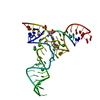 8zauC C: citing same article ( |
|---|---|
| Similar structure data |
-Links
 PDBj
PDBj


- Assembly
| Deposited unit | 
| ||||||||
|---|---|---|---|---|---|---|---|---|---|
| 1 | 
| ||||||||
| Unit cell |
|
-Components
| #1: RNA chain | Mass: 13256.007 Da / Num. of mol.: 2 / Source method: obtained synthetically / Source: (synth.) Homo (humans)#2: Chemical | ChemComp-NA /   Mass: 22.990 Da / Num. of mol.: 5 / Source method: obtained synthetically / Formula: Na / Feature type: SUBJECT OF INVESTIGATION Mass: 22.990 Da / Num. of mol.: 5 / Source method: obtained synthetically / Formula: Na / Feature type: SUBJECT OF INVESTIGATION#3: Water | ChemComp-HOH / |  Mass: 18.015 Da / Num. of mol.: 4 / Source method: isolated from a natural source / Formula: H2O Mass: 18.015 Da / Num. of mol.: 4 / Source method: isolated from a natural source / Formula: H2OHas ligand of interest | Y | Has protein modification | N | |
|---|
-Experimental details
-Experiment
| Experiment | Method: X-RAY DIFFRACTION / Number of used crystals: 1 |
|---|
- Sample preparation
| Crystal | Density Matthews: 6.22 Å3/Da / Density % sol: 80.21 % |
|---|---|
| Crystal grow | Temperature: 291 K / Method: vapor diffusion, hanging drop Details: 0.02 M Calcium Chloride dihydrate 0.1 M Sodium Acetate trihydrate pH 4.6 30% v/v 2-Methyl-2,4-pentanediol |
-Data collection
| Diffraction | Mean temperature: 100 K / Serial crystal experiment: N |
|---|---|
| Diffraction source | Source: SYNCHROTRON / Site: SSRF / Beamline: BL19U1 / Wavelength: 0.97853 Å |
| Detector | Type: DECTRIS PILATUS3 6M / Detector: PIXEL / Date: Nov 7, 2021 |
| Radiation | Protocol: SINGLE WAVELENGTH / Monochromatic (M) / Laue (L): M / Scattering type: x-ray |
| Radiation wavelength | Wavelength: 0.97853 Å / Relative weight: 1 |
| Reflection | Resolution: 3.86→80.75 Å / Num. obs: 6885 / % possible obs: 98.2 % / Redundancy: 18 % / CC1/2: 0.999 / Net I/σ(I): 22 |
| Reflection shell | Resolution: 3.86→4.06 Å / Num. unique obs: 863 / CC1/2: 0.976 |
- Processing
| Software |
| ||||||||||||||||||||||||||||||||||||||||
|---|---|---|---|---|---|---|---|---|---|---|---|---|---|---|---|---|---|---|---|---|---|---|---|---|---|---|---|---|---|---|---|---|---|---|---|---|---|---|---|---|---|
| Refinement | Method to determine structure: MOLECULAR REPLACEMENT / Resolution: 3.86→36.67 Å / SU ML: 0.5 / Cross valid method: FREE R-VALUE / σ(F): 1.35 / Phase error: 45.16 / Stereochemistry target values: ML
| ||||||||||||||||||||||||||||||||||||||||
| Solvent computation | Shrinkage radii: 0.9 Å / VDW probe radii: 1.11 Å / Solvent model: FLAT BULK SOLVENT MODEL | ||||||||||||||||||||||||||||||||||||||||
| Refinement step | Cycle: LAST / Resolution: 3.86→36.67 Å
| ||||||||||||||||||||||||||||||||||||||||
| Refine LS restraints |
| ||||||||||||||||||||||||||||||||||||||||
| LS refinement shell |
| ||||||||||||||||||||||||||||||||||||||||
| Refinement TLS params. | Method: refined / Origin x: -36.6508 Å / Origin y: 7.9415 Å / Origin z: -36.6024 Å
| ||||||||||||||||||||||||||||||||||||||||
| Refinement TLS group | Selection details: all |
