 Movie
Movie Controller

[English] 日本語
 Yorodumi
- PDB-8z2b: Crystal structure of apo Aspergillus terreus glutamate dehydrogen... -
+ Open data

- Basic information
| Entry | Database: PDB / ID: 8z2b | ||||||
|---|---|---|---|---|---|---|---|
| Title | Crystal structure of apo Aspergillus terreus glutamate dehydrogenase (AtGDH) in the partially closed conformation (form II) | ||||||
 Components Components | Glutamate dehydrogenase | ||||||
 Keywords Keywords | OXIDOREDUCTASE / glutamate dehydrogenase / allostery / cooperativity / Aspergillus / cryo-EM / domain dynamics | ||||||
| Function / homology |  Function and homology information Function and homology informationglutamate biosynthetic process / glutamate dehydrogenase (NADP+) activity / nucleotide binding / cytosol Similarity search - Function | ||||||
| Biological species |  | ||||||
| Method |  X-RAY DIFFRACTION / MOLECULAR REPLACEMENT / Resolution: 2.85 Å X-RAY DIFFRACTION / MOLECULAR REPLACEMENT / Resolution: 2.85 Å | ||||||
 Authors Authors | Godsora, B.K.J. / Bhaumik, P. | ||||||
| Funding support |  India, 1items India, 1items
| ||||||
 Citation Citation | Journal: Protein Sci / Year: 2025 Title: Conformational flexibility associated with remote residues regulates the kinetic properties of glutamate dehydrogenase. Authors: Barsa Kanchan Jyotshna Godsora / Parijat Das / Prasoon Kumar Mishra / Anjali Sairaman / Sandip Kaledhonkar / Narayan S Punekar / Prasenjit Bhaumik / Abstract: Glutamate dehydrogenase (GDH) is a pivotal metabolic enzyme in all living organisms, and some of the GDHs exhibit substrate-dependent homotropic cooperativity. However, the mode of allosteric ...Glutamate dehydrogenase (GDH) is a pivotal metabolic enzyme in all living organisms, and some of the GDHs exhibit substrate-dependent homotropic cooperativity. However, the mode of allosteric communication during the homotropic effect in GDHs remains poorly understood. In this study, we examined two homologous GDHs, Aspergillus niger GDH (AnGDH) and Aspergillus terreus GDH (AtGDH), with differing substrate utilization kinetics to uncover the factors driving their distinct behavior. We report the crystal structures and first-ever cryo-EM structures of apo- AtGDH and AnGDH that captured arrays of conformational ensembles. A wider mouth opening (~ 21 Å) is observed for the cooperative AnGDH as compared to the non-cooperative AtGDH (~17 Å) in their apo states. A network of interactions related to the substitutions in Domain II influence structural flexibility in these GDHs. Remarkably, we have identified a distant substitution (R246 to S) in Domain II, as a part of this network, which can impact the mouth opening and converts non-cooperative AtGDH into a cooperative enzyme. Our study demonstrates that remote residues can influence structural and kinetic properties in homologous GDHs. | ||||||
| History |
|
- Structure visualization
| Structure viewer | Molecule: MolmilJmol/JSmol |
|---|
- Downloads & links
-Download
| PDBx/mmCIF format | 8z2b.cif.gz | 523.5 KB | Display | PDBx/mmCIF format |
|---|---|---|---|---|
| PDB format | pdb8z2b.ent.gz | 420.1 KB | Display | PDB format |
| PDBx/mmJSON format | 8z2b.json.gz | Tree view | PDBx/mmJSON format | |
| Others |  Other downloads Other downloads |
-Validation report
| Summary document | 8z2b_validation.pdf.gz | 496.8 KB | Display | wwPDB validaton report |
|---|---|---|---|---|
| Full document | 8z2b_full_validation.pdf.gz | 583.9 KB | Display | |
| Data in XML | 8z2b_validation.xml.gz | 112.3 KB | Display | |
| Data in CIF | 8z2b_validation.cif.gz | 142 KB | Display | |
| Arichive directory | https://data.pdbj.org/pub/pdb/validation_reports/z2/8z2bftp://data.pdbj.org/pub/pdb/validation_reports/z2/8z2b | HTTPS FTP |
-Related structure data
| Related structure data |  8z1cC  8z1mC  8z1nC  8z1oC  8z29C  8z2aC  8z2cC  8z2fC 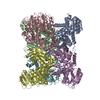 5xviS S: Starting model for refinement C: citing same article ( |
|---|---|
| Similar structure data |


-Links
 PDBj
PDBj- Assembly
| Deposited unit | 
| ||||||||||||||||||||||||||||||||||||||||||||||||||||||||||||||||||||||||||||||||||||||||||||||||||||||||||||||||||||||||||||||||||||||||||||||||||||||||||||||||||||||||||||||||||||||||||||||||||||||||||||||||||||||||||
|---|---|---|---|---|---|---|---|---|---|---|---|---|---|---|---|---|---|---|---|---|---|---|---|---|---|---|---|---|---|---|---|---|---|---|---|---|---|---|---|---|---|---|---|---|---|---|---|---|---|---|---|---|---|---|---|---|---|---|---|---|---|---|---|---|---|---|---|---|---|---|---|---|---|---|---|---|---|---|---|---|---|---|---|---|---|---|---|---|---|---|---|---|---|---|---|---|---|---|---|---|---|---|---|---|---|---|---|---|---|---|---|---|---|---|---|---|---|---|---|---|---|---|---|---|---|---|---|---|---|---|---|---|---|---|---|---|---|---|---|---|---|---|---|---|---|---|---|---|---|---|---|---|---|---|---|---|---|---|---|---|---|---|---|---|---|---|---|---|---|---|---|---|---|---|---|---|---|---|---|---|---|---|---|---|---|---|---|---|---|---|---|---|---|---|---|---|---|---|---|---|---|---|---|---|---|---|---|---|---|---|---|---|---|---|---|---|---|---|---|
| 1 |
| ||||||||||||||||||||||||||||||||||||||||||||||||||||||||||||||||||||||||||||||||||||||||||||||||||||||||||||||||||||||||||||||||||||||||||||||||||||||||||||||||||||||||||||||||||||||||||||||||||||||||||||||||||||||||||
| Unit cell |
| ||||||||||||||||||||||||||||||||||||||||||||||||||||||||||||||||||||||||||||||||||||||||||||||||||||||||||||||||||||||||||||||||||||||||||||||||||||||||||||||||||||||||||||||||||||||||||||||||||||||||||||||||||||||||||
| Noncrystallographic symmetry (NCS) | NCS domain:
NCS domain segments: Beg auth comp-ID: MET / Beg label comp-ID: MET / End auth comp-ID: TRP / End label comp-ID: TRP / Auth asym-ID: A / Label asym-ID: A / Auth seq-ID: 1 - 460 / Label seq-ID: 1 - 460
NCS ensembles :
|
-Components
| #1: Protein | Mass: 49247.438 Da / Num. of mol.: 6 Source method: isolated from a genetically manipulated source Source: (gene. exp.)  #2: Water | ChemComp-HOH / |  Mass: 18.015 Da / Num. of mol.: 23 / Source method: isolated from a natural source / Formula: H2O Mass: 18.015 Da / Num. of mol.: 23 / Source method: isolated from a natural source / Formula: H2OHas protein modification | N | |
|---|
-Experimental details
-Experiment
| Experiment | Method: X-RAY DIFFRACTION / Number of used crystals: 1 |
|---|
- Sample preparation
| Crystal | Density Matthews: 2.74 Å3/Da / Density % sol: 55.19 % |
|---|---|
| Crystal grow | Temperature: 295 K / Method: vapor diffusion, hanging drop / Details: 0.2 M MgCl2, 0.1 M Tris pH 7, and 10% PEG 8000 |
-Data collection
| Diffraction | Mean temperature: 100 K / Serial crystal experiment: N | ||||||||||||||||||||||||||||||
|---|---|---|---|---|---|---|---|---|---|---|---|---|---|---|---|---|---|---|---|---|---|---|---|---|---|---|---|---|---|---|---|
| Diffraction source | Source: ROTATING ANODE / Type: RIGAKU MICROMAX-007 HF / Wavelength: 1.5418 Å | ||||||||||||||||||||||||||||||
| Detector | Type: RIGAKU RAXIS IV++ / Detector: IMAGE PLATE / Date: Aug 17, 2017 | ||||||||||||||||||||||||||||||
| Radiation | Protocol: SINGLE WAVELENGTH / Monochromatic (M) / Laue (L): M / Scattering type: x-ray | ||||||||||||||||||||||||||||||
| Radiation wavelength | Wavelength: 1.5418 Å / Relative weight: 1 | ||||||||||||||||||||||||||||||
| Reflection twin |
| ||||||||||||||||||||||||||||||
| Reflection | Resolution: 2.85→30 Å / Num. obs: 73339 / % possible obs: 99.9 % / Redundancy: 5.65 % / CC1/2: 0.98 / Net I/σ(I): 9.3 | ||||||||||||||||||||||||||||||
| Reflection shell | Resolution: 2.85→2.95 Å / Num. unique obs: 7224 / CC1/2: 0.61 |
- Processing
| Software |
| ||||||||||||||||||||||||||||||||||||||||||||||||||||||||||||||||||||||||||||||||||||||||||||||||||||||||||||||||||||||||||||||||||||||||||||||||||||||||||||||||||||||||||||||||||||||
|---|---|---|---|---|---|---|---|---|---|---|---|---|---|---|---|---|---|---|---|---|---|---|---|---|---|---|---|---|---|---|---|---|---|---|---|---|---|---|---|---|---|---|---|---|---|---|---|---|---|---|---|---|---|---|---|---|---|---|---|---|---|---|---|---|---|---|---|---|---|---|---|---|---|---|---|---|---|---|---|---|---|---|---|---|---|---|---|---|---|---|---|---|---|---|---|---|---|---|---|---|---|---|---|---|---|---|---|---|---|---|---|---|---|---|---|---|---|---|---|---|---|---|---|---|---|---|---|---|---|---|---|---|---|---|---|---|---|---|---|---|---|---|---|---|---|---|---|---|---|---|---|---|---|---|---|---|---|---|---|---|---|---|---|---|---|---|---|---|---|---|---|---|---|---|---|---|---|---|---|---|---|---|---|
| Refinement | Method to determine structure: MOLECULAR REPLACEMENT Starting model: 5XVI Resolution: 2.85→29.89 Å / Cor.coef. Fo:Fc: 0.876 / Cor.coef. Fo:Fc free: 0.773 / SU B: 12.687 / SU ML: 0.266 / Cross valid method: FREE R-VALUE / ESU R Free: 0.09 Details: Hydrogens have been added in their riding positions
| ||||||||||||||||||||||||||||||||||||||||||||||||||||||||||||||||||||||||||||||||||||||||||||||||||||||||||||||||||||||||||||||||||||||||||||||||||||||||||||||||||||||||||||||||||||||
| Solvent computation | Ion probe radii: 0.8 Å / Shrinkage radii: 0.8 Å / VDW probe radii: 1.2 Å / Solvent model: MASK BULK SOLVENT | ||||||||||||||||||||||||||||||||||||||||||||||||||||||||||||||||||||||||||||||||||||||||||||||||||||||||||||||||||||||||||||||||||||||||||||||||||||||||||||||||||||||||||||||||||||||
| Displacement parameters | Biso mean: 47.117 Å2
| ||||||||||||||||||||||||||||||||||||||||||||||||||||||||||||||||||||||||||||||||||||||||||||||||||||||||||||||||||||||||||||||||||||||||||||||||||||||||||||||||||||||||||||||||||||||
| Refinement step | Cycle: LAST / Resolution: 2.85→29.89 Å
| ||||||||||||||||||||||||||||||||||||||||||||||||||||||||||||||||||||||||||||||||||||||||||||||||||||||||||||||||||||||||||||||||||||||||||||||||||||||||||||||||||||||||||||||||||||||
| Refine LS restraints |
|