 Movie
Movie Controller


+ Open data

- Basic information
| Entry | Database: PDB / ID: 8ypd | ||||||
|---|---|---|---|---|---|---|---|
| Title | Cryo-EM structure of the LH1 complex from Allochromatium tepidum | ||||||
 Components Components |
| ||||||
 Keywords Keywords | PHOTOSYNTHESIS / LH1 COMPLEX | ||||||
| Function / homology |  Function and homology information Function and homology informationplasma membrane light-harvesting complex / bacteriochlorophyll binding / photosynthesis, light reaction / metal ion binding / plasma membrane Similarity search - Function | ||||||
| Biological species |  Allochromatium tepidum (bacteria) Allochromatium tepidum (bacteria) | ||||||
| Method | ELECTRON MICROSCOPY / single particle reconstruction / cryo EM / Resolution: 2.78 Å | ||||||
 Authors Authors | Wang, G.-L. / Sun, S. / Yu, L.-J. | ||||||
| Funding support |  China, 1items China, 1items
| ||||||
 Citation Citation | Journal: Biomolecules / Year: 2025 Title: Probing the Dual Role of Ca in the LH1-RC Complex by Constructing and Analyzing Ca-Bound and Ca-Free LH1 Complexes. Authors: Mei-Juan Zou / Shuai Sun / Guang-Lei Wang / Yi-Hao Yan / Wei Ji / Zheng-Yu Wang-Otomo / Michael T Madigan / Long-Jiang Yu /   Abstract: The genome of the mildly thermophilic hot spring purple sulfur bacterium, (.) , contains a multigene family that encodes a series of α- and β-polypeptides, collectively forming a heterogeneous ...The genome of the mildly thermophilic hot spring purple sulfur bacterium, (.) , contains a multigene family that encodes a series of α- and β-polypeptides, collectively forming a heterogeneous light-harvesting 1 (LH1) complex. The LH1, therefore, offers a unique model for studying an intermediate phenotype between phototrophic thermophilic and mesophilic bacteria, particularly regarding their LH1 transition and moderately enhanced thermal stability. Of the 16 α-polypeptides in the LH1, six α1 bind Ca to connect with β1- or β3-polypeptides in specific Ca-binding sites. Here, we use the purple bacterium strain H2 as a host to express Ca-bound and Ca-free LH1-only complexes composed of α- and β-polypeptides that either contain or lack the calcium-binding motif WxxDxI; purified preparations of each complex were then used to test how Ca affects their thermostability and spectral features. The cryo-EM structures of both complexes were closed circular rings consisting of 14 αβ-polypeptides. The absorption maximum of Ca-bound LH1 (α1/β1 and α1/β3) was at 894 nm, while that of Ca-free (α2/β1) was at 888 nm, indicating that Ca imparts a transition of 6 nm. Crucially for the ecological success of , Ca-bound LH1 complexes were more thermostable than Ca-free complexes, indicating that calcium plays at least two major roles in photosynthesis by -improving photocomplex stability and modifying its spectrum. | ||||||
| History |
|
- Structure visualization
| Structure viewer | Molecule: MolmilJmol/JSmol |
|---|
- Downloads & links
-Download
| PDBx/mmCIF format | 8ypd.cif.gz | 263.7 KB | Display | PDBx/mmCIF format |
|---|---|---|---|---|
| PDB format | pdb8ypd.ent.gz | 233 KB | Display | PDB format |
| PDBx/mmJSON format | 8ypd.json.gz | Tree view | PDBx/mmJSON format | |
| Others |  Other downloads Other downloads |
-Validation report
| Arichive directory | https://data.pdbj.org/pub/pdb/validation_reports/yp/8ypdftp://data.pdbj.org/pub/pdb/validation_reports/yp/8ypd | HTTPS FTP |
|---|
-Related structure data
| Related structure data | 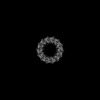 39477MC  8ypbC C: citing same article ( M: map data used to model this data |
|---|---|
| Similar structure data |

-Links
 PDBj
PDBj- Assembly
| Deposited unit | 
|
|---|---|
| 1 |
|
-Components
| #1: Protein/peptide | Mass: 5269.103 Da / Num. of mol.: 14 Source method: isolated from a genetically manipulated source Source: (gene. exp.) Allochromatium tepidum (bacteria) / Gene: pufBProduction host: Rhodospirillum rubrum ATCC 11170 (bacteria)References: UniProt: O82943 #2: Protein/peptide | Mass: 5108.121 Da / Num. of mol.: 14 Source method: isolated from a genetically manipulated source Source: (gene. exp.) Allochromatium tepidum (bacteria)Production host: Rhodospirillum rubrum ATCC 11170 (bacteria)#3: Chemical | ChemComp-BCL /   Mass: 911.504 Da / Num. of mol.: 28 / Source method: obtained synthetically / Formula: C55H74MgN4O6 / Feature type: SUBJECT OF INVESTIGATION Mass: 911.504 Da / Num. of mol.: 28 / Source method: obtained synthetically / Formula: C55H74MgN4O6 / Feature type: SUBJECT OF INVESTIGATION#4: Chemical | ChemComp-CRT /   Mass: 596.925 Da / Num. of mol.: 14 / Source method: obtained synthetically / Formula: C42H60O2 / Feature type: SUBJECT OF INVESTIGATION Mass: 596.925 Da / Num. of mol.: 14 / Source method: obtained synthetically / Formula: C42H60O2 / Feature type: SUBJECT OF INVESTIGATIONHas ligand of interest | Y | Has protein modification | N | |
|---|
-Experimental details
-Experiment
| Experiment | Method: ELECTRON MICROSCOPY |
|---|---|
| EM experiment | Aggregation state: PARTICLE / 3D reconstruction method: single particle reconstruction |
- Sample preparation
| Component | Name: LH1 COMPLEX / Type: COMPLEX / Entity ID: #1-#2 / Source: RECOMBINANT |
|---|---|
| Molecular weight | Experimental value: NO |
| Source (natural) | Organism: Allochromatium tepidum (bacteria) |
| Source (recombinant) | Organism: Rhodospirillum rubrum ATCC 11170 (bacteria) |
| Buffer solution | pH: 7.5 |
| Specimen | Embedding applied: NO / Shadowing applied: NO / Staining applied: NO / Vitrification applied: YES |
| Vitrification | Cryogen name: ETHANE |
- Electron microscopy imaging
| Experimental equipment |  Model: Titan Krios / Image courtesy: FEI Company |
|---|---|
| Microscopy | Model: TFS KRIOS |
| Electron gun | Electron source:  FIELD EMISSION GUN / Accelerating voltage: 300 kV / Illumination mode: FLOOD BEAM FIELD EMISSION GUN / Accelerating voltage: 300 kV / Illumination mode: FLOOD BEAM |
| Electron lens | Mode: BRIGHT FIELD / Nominal defocus max: 2200 nm / Nominal defocus min: 800 nm |
| Image recording | Electron dose: 60 e/Å2 / Film or detector model: GATAN K3 (6k x 4k) |
- Processing
| EM software | Name: PHENIX / Version: 1.19.2_4158: / Category: model refinement | ||||||||||||||||||||||||
|---|---|---|---|---|---|---|---|---|---|---|---|---|---|---|---|---|---|---|---|---|---|---|---|---|---|
| CTF correction | Type: PHASE FLIPPING ONLY | ||||||||||||||||||||||||
| 3D reconstruction | Resolution: 2.78 Å / Resolution method: FSC 0.143 CUT-OFF / Num. of particles: 199061 / Symmetry type: POINT | ||||||||||||||||||||||||
| Refine LS restraints |
|
