 Movie
Movie Controller


+ Open data

- Basic information
| Entry | Database: PDB / ID: 8yh2 | |||||||||||||||||||||
|---|---|---|---|---|---|---|---|---|---|---|---|---|---|---|---|---|---|---|---|---|---|---|
| Title | A3R-Gi complex bound to adenosine | |||||||||||||||||||||
 Components Components |
| |||||||||||||||||||||
 Keywords Keywords | SIGNALING PROTEIN / GPCR / adenosine receptor / complex / G protein | |||||||||||||||||||||
| Function / homology |  Function and homology information Function and homology informationOlfactory Signaling Pathway / Sensory perception of sweet, bitter, and umami (glutamate) taste / Synthesis, secretion, and inactivation of Glucagon-like Peptide-1 (GLP-1) / Activation of the phototransduction cascade / G protein-coupled adenosine receptor activity / Activation of G protein gated Potassium channels / G-protein activation / G beta:gamma signalling through PI3Kgamma / Prostacyclin signalling through prostacyclin receptor / G beta:gamma signalling through PLC beta ...Olfactory Signaling Pathway / Sensory perception of sweet, bitter, and umami (glutamate) taste / Synthesis, secretion, and inactivation of Glucagon-like Peptide-1 (GLP-1) / Activation of the phototransduction cascade / G protein-coupled adenosine receptor activity / Activation of G protein gated Potassium channels / G-protein activation / G beta:gamma signalling through PI3Kgamma / Prostacyclin signalling through prostacyclin receptor / G beta:gamma signalling through PLC beta / ADP signalling through P2Y purinoceptor 1 / Thromboxane signalling through TP receptor / Presynaptic function of Kainate receptors / G beta:gamma signalling through CDC42 / Inhibition of voltage gated Ca2+ channels via Gbeta/gamma subunits / G alpha (12/13) signalling events / Glucagon-type ligand receptors / G beta:gamma signalling through BTK / ADP signalling through P2Y purinoceptor 12 / Adrenaline,noradrenaline inhibits insulin secretion / Cooperation of PDCL (PhLP1) and TRiC/CCT in G-protein beta folding / Ca2+ pathway / G alpha (z) signalling events / Thrombin signalling through proteinase activated receptors (PARs) / Extra-nuclear estrogen signaling / G alpha (s) signalling events / G alpha (q) signalling events / Glucagon-like Peptide-1 (GLP1) regulates insulin secretion / G alpha (i) signalling events / High laminar flow shear stress activates signaling by PIEZO1 and PECAM1:CDH5:KDR in endothelial cells / Vasopressin regulates renal water homeostasis via Aquaporins / regulation of eating behavior / adenylate cyclase inhibitor activity / positive regulation of protein localization to cell cortex / T cell migration / positive regulation of relaxation of smooth muscle / Adenylate cyclase inhibitory pathway / D2 dopamine receptor binding / adenylate cyclase-inhibiting serotonin receptor signaling pathway / G protein-coupled serotonin receptor binding / viral budding from plasma membrane / cellular response to forskolin / bioluminescence / mast cell degranulation / regulation of mitotic spindle organization / chemokine-mediated signaling pathway / generation of precursor metabolites and energy / Regulation of insulin secretion / neuropeptide signaling pathway / response to prostaglandin E / positive regulation of cholesterol biosynthetic process / G protein-coupled receptor binding / response to peptide hormone / G-protein beta/gamma-subunit complex binding / adenylate cyclase-modulating G protein-coupled receptor signaling pathway / adenylate cyclase-inhibiting G protein-coupled receptor signaling pathway / G beta:gamma signalling through PLC beta / Presynaptic function of Kainate receptors / Thromboxane signalling through TP receptor / Activation of G protein gated Potassium channels / Inhibition of voltage gated Ca2+ channels via Gbeta/gamma subunits / G-protein activation / Glucagon signaling in metabolic regulation / G beta:gamma signalling through CDC42 / Prostacyclin signalling through prostacyclin receptor / G beta:gamma signalling through BTK / photoreceptor disc membrane / GDP binding / ADP signalling through P2Y purinoceptor 12 / Glucagon-type ligand receptors / Adrenaline,noradrenaline inhibits insulin secretion / Vasopressin regulates renal water homeostasis via Aquaporins / Glucagon-like Peptide-1 (GLP1) regulates insulin secretion / G alpha (z) signalling events / cellular response to catecholamine stimulus / ADP signalling through P2Y purinoceptor 1 / G beta:gamma signalling through PI3Kgamma / ADORA2B mediated anti-inflammatory cytokines production / adenylate cyclase-activating dopamine receptor signaling pathway / Cooperation of PDCL (PhLP1) and TRiC/CCT in G-protein beta folding / GPER1 signaling / cellular response to prostaglandin E stimulus / heterotrimeric G-protein complex / G alpha (12/13) signalling events / G-protein beta-subunit binding / sensory perception of taste / Thrombin signalling through proteinase activated receptors (PARs) / signaling receptor complex adaptor activity / adenylate cyclase-activating G protein-coupled receptor signaling pathway / retina development in camera-type eye / fibroblast proliferation / GTPase binding / G protein activity / midbody / Ca2+ pathway / cell cortex / clathrin-dependent endocytosis of virus by host cell / High laminar flow shear stress activates signaling by PIEZO1 and PECAM1:CDH5:KDR in endothelial cells / G alpha (i) signalling events / G alpha (s) signalling events Similarity search - Function | |||||||||||||||||||||
| Biological species |   Homo sapiens (human) Homo sapiens (human)  Influenza A virusHuman orthopneumovirus Influenza A virusHuman orthopneumovirus | |||||||||||||||||||||
| Method | ELECTRON MICROSCOPY / single particle reconstruction / cryo EM / Resolution: 3.27 Å | |||||||||||||||||||||
 Authors Authors | Oshima, H.S. / Shihoya, W. / Nureki, O. | |||||||||||||||||||||
| Funding support |  Japan, 1items Japan, 1items
| |||||||||||||||||||||
 Citation Citation | Journal: Nat Commun / Year: 2024 Title: Structural insights into the agonist selectivity of the adenosine A receptor. Authors: Hidetaka S Oshima / Akiko Ogawa / Fumiya K Sano / Hiroaki Akasaka / Tomoyoshi Kawakami / Aika Iwama / Hiroyuki H Okamoto / Chisae Nagiri / Fan-Yan Wei / Wataru Shihoya / Osamu Nureki / Abstract: Adenosine receptors play pivotal roles in physiological processes. Adenosine A receptor (AR), the most recently identified adenosine receptor, is expressed in various tissues, exhibiting important ...Adenosine receptors play pivotal roles in physiological processes. Adenosine A receptor (AR), the most recently identified adenosine receptor, is expressed in various tissues, exhibiting important roles in neuron, heart, and immune cells, and is often overexpressed in tumors, highlighting the therapeutic potential of AR-selective agents. Recently, we identified RNA-derived N-methyladenosine (mA) as an endogenous agonist for AR, suggesting the relationship between RNA-derived modified adenosine and AR. Despite extensive studies on the other adenosine receptors, the selectivity mechanism of AR, especially for AR-selective agonists such as mA and namodenoson, remained elusive. Here, we identify tRNA-derived N-isopentenyl adenosine (iA) as an AR-selective ligand via screening of modified nucleosides against the adenosine receptors. Like mA, iA is found in the human body and may be an endogenous AR ligand. Our cryo-EM analyses elucidate the AR-G complexes bound to adenosine, 5'-N-ethylcarboxamidoadenosine (NECA), mA, iA, and namodenoson at overall resolutions of 3.27 Å (adenosine), 2.86 Å (NECA), 3.19 Å (mA), 3.28 Å (iA), and 3.20 Å (namodenoson), suggesting the selectivity and activation mechanism of AR. We further conduct structure-guided engineering of mA-insensitive AR, which may aid future research targeting mA and AR, providing a molecular basis for future drug discovery. | |||||||||||||||||||||
| History |
|
- Structure visualization
| Structure viewer | Molecule: MolmilJmol/JSmol |
|---|
- Downloads & links
-Download
| PDBx/mmCIF format | 8yh2.cif.gz | 231.5 KB | Display | PDBx/mmCIF format |
|---|---|---|---|---|
| PDB format | pdb8yh2.ent.gz | 166.4 KB | Display | PDB format |
| PDBx/mmJSON format | 8yh2.json.gz | Tree view | PDBx/mmJSON format | |
| Others |  Other downloads Other downloads |
-Validation report
| Arichive directory | https://data.pdbj.org/pub/pdb/validation_reports/yh/8yh2ftp://data.pdbj.org/pub/pdb/validation_reports/yh/8yh2 | HTTPS FTP |
|---|
-Related structure data
| Related structure data |  39279MC 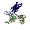 8yh0C  8yh3C  8yh5C 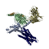 8yh6C M: map data used to model this data C: citing same article ( |
|---|---|
| Similar structure data |


-Links
 PDBj
PDBj


- Assembly
| Deposited unit | 
|
|---|---|
| 1 |
|
-Components
| #1: Protein | Mass: 40586.332 Da / Num. of mol.: 1 Source method: isolated from a genetically manipulated source Source: (gene. exp.) Homo sapiens (human) / References: UniProt: P62871 | ||||||||||
|---|---|---|---|---|---|---|---|---|---|---|---|
| #2: Protein | Mass: 48846.695 Da / Num. of mol.: 2 Source method: isolated from a genetically manipulated source Source: (gene. exp.) Homo sapiens (human) / Gene: GNG2, GNAI1 / Production host: Homo sapiens (human) / References: UniProt: P59768, UniProt: P63096#3: Protein | | Mass: 88315.891 Da / Num. of mol.: 1 Source method: isolated from a genetically manipulated source Source: (gene. exp.) Influenza A virus (A/Victoria/3/1975(H3N2)), (gene. exp.) Human orthopneumovirusStrain: A/Victoria/3/1975(H3N2) / Gene: HA, ADORA3 / Production host: Homo sapiens (human)References: UniProt: P03435, UniProt: W5QED6, UniProt: A0A5P9VSM6 #4: Antibody | | Mass: 27720.795 Da / Num. of mol.: 1 Source method: isolated from a genetically manipulated source Source: (gene. exp.) Homo sapiens (human)#5: Chemical | ChemComp-ADN / |   Mass: 267.241 Da / Num. of mol.: 1 / Source method: obtained synthetically / Formula: C10H13N5O4 / Feature type: SUBJECT OF INVESTIGATION Mass: 267.241 Da / Num. of mol.: 1 / Source method: obtained synthetically / Formula: C10H13N5O4 / Feature type: SUBJECT OF INVESTIGATIONHas ligand of interest | Y | Has protein modification | Y | |
-Experimental details
-Experiment
| Experiment | Method: ELECTRON MICROSCOPY |
|---|---|
| EM experiment | Aggregation state: PARTICLE / 3D reconstruction method: single particle reconstruction |
- Sample preparation
| Component | Name: A3R-Gi complex bound to adenosine / Type: COMPLEX / Entity ID: #1, #3, #2, #4 / Source: RECOMBINANT |
|---|---|
| Source (natural) | Organism: |
| Source (recombinant) | Organism: Homo sapiens (human) |
| Buffer solution | pH: 8 |
| Specimen | Embedding applied: NO / Shadowing applied: NO / Staining applied: NO / Vitrification applied: YES |
| Vitrification | Cryogen name: ETHANE |
- Electron microscopy imaging
| Experimental equipment |  Model: Titan Krios / Image courtesy: FEI Company |
|---|---|
| Microscopy | Model: FEI TITAN KRIOS |
| Electron gun | Electron source:  FIELD EMISSION GUN / Accelerating voltage: 300 kV / Illumination mode: FLOOD BEAM FIELD EMISSION GUN / Accelerating voltage: 300 kV / Illumination mode: FLOOD BEAM |
| Electron lens | Mode: BRIGHT FIELD / Nominal defocus max: 1600 nm / Nominal defocus min: 800 nm |
| Image recording | Electron dose: 50 e/Å2 / Film or detector model: GATAN K3 (6k x 4k) |
- Processing
| EM software | Name: PHENIX / Category: model refinement |
|---|---|
| CTF correction | Type: PHASE FLIPPING AND AMPLITUDE CORRECTION |
| 3D reconstruction | Resolution: 3.27 Å / Resolution method: FSC 0.143 CUT-OFF / Num. of particles: 160357 / Symmetry type: POINT |
