Movie
Movie Controller

[English] 日本語
 Yorodumi
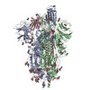
- PDB-8ydx: Cryo-EM structure of SARS-CoV-2 spike ectodomain (HexaPro, Omicro... -
+ Open data

- Basic information
| Entry | Database: PDB / ID: 8ydx | ||||||||||||||||||
|---|---|---|---|---|---|---|---|---|---|---|---|---|---|---|---|---|---|---|---|
| Title | Cryo-EM structure of SARS-CoV-2 spike ectodomain (HexaPro, Omicron BA.2 variant) in complex with CeSPIACE | ||||||||||||||||||
 Components Components |
| ||||||||||||||||||
 Keywords Keywords | VIRAL PROTEIN/INHIBITOR / Spike protein / VIRAL PROTEIN-INHIBITOR COMPLEX | ||||||||||||||||||
| Function / homology |  Function and homology information Function and homology informationsymbiont-mediated disruption of host tissue / Maturation of spike protein / Translation of Structural Proteins / Virion Assembly and Release / host cell surface / host extracellular region / symbiont-mediated-mediated suppression of host tetherin activity / Induction of Cell-Cell Fusion / structural constituent of virion / positive regulation of viral entry into host cell ...symbiont-mediated disruption of host tissue / Maturation of spike protein / Translation of Structural Proteins / Virion Assembly and Release / host cell surface / host extracellular region / symbiont-mediated-mediated suppression of host tetherin activity / Induction of Cell-Cell Fusion / structural constituent of virion / positive regulation of viral entry into host cell / membrane fusion / host cell endoplasmic reticulum-Golgi intermediate compartment membrane / Attachment and Entry / entry receptor-mediated virion attachment to host cell / receptor-mediated virion attachment to host cell / host cell surface receptor binding / symbiont-mediated suppression of host innate immune response / endocytosis involved in viral entry into host cell / receptor ligand activity / fusion of virus membrane with host plasma membrane / fusion of virus membrane with host endosome membrane / viral envelope / symbiont entry into host cell / virion attachment to host cell / host cell plasma membrane / SARS-CoV-2 activates/modulates innate and adaptive immune responses / virion membrane / membrane / identical protein binding / plasma membrane Similarity search - Function | ||||||||||||||||||
| Biological species |   Severe acute respiratory syndrome coronavirus 2 Severe acute respiratory syndrome coronavirus 2synthetic construct (others) | ||||||||||||||||||
| Method | ELECTRON MICROSCOPY / single particle reconstruction / cryo EM / Resolution: 4.9 Å | ||||||||||||||||||
 Authors Authors | Suzuki, H. / Nakamura, S. / Fujiyoshi, Y. | ||||||||||||||||||
| Funding support |  Japan, 5items Japan, 5items
| ||||||||||||||||||
 Citation Citation | Journal: Proc Natl Acad Sci U S A / Year: 2025 Title: Structure-guided engineering of a mutation-tolerant inhibitor peptide against variable SARS-CoV-2 spikes. Authors: Shun Nakamura / Yukihiro Tanimura / Risa Nomura / Hiroshi Suzuki / Kouki Nishikawa / Akiko Kamegawa / Nobutaka Numoto / Atsushi Tanaka / Shigeru Kawabata / Shoichi Sakaguchi / Akino Emi / ...Authors: Shun Nakamura / Yukihiro Tanimura / Risa Nomura / Hiroshi Suzuki / Kouki Nishikawa / Akiko Kamegawa / Nobutaka Numoto / Atsushi Tanaka / Shigeru Kawabata / Shoichi Sakaguchi / Akino Emi / Youichi Suzuki / Yoshinori Fujiyoshi / Abstract: Pathogen mutations present an inevitable and challenging problem for therapeutics and the development of mutation-tolerant anti-infective drugs to strengthen global health and combat evolving ...Pathogen mutations present an inevitable and challenging problem for therapeutics and the development of mutation-tolerant anti-infective drugs to strengthen global health and combat evolving pathogens is urgently needed. While spike proteins on viral surfaces are attractive targets for preventing viral entry, they mutate frequently, making it difficult to develop effective therapeutics. Here, we used a structure-guided strategy to engineer an inhibitor peptide against the SARS-CoV-2 spike, called CeSPIACE, with mutation-tolerant and potent binding ability against all variants to enhance affinity for the invariant architecture of the receptor-binding domain (RBD). High-resolution structures of the peptide complexed with mutant RBDs revealed a mechanism of mutation-tolerant inhibition. CeSPIACE bound major mutant RBDs with picomolar affinity and inhibited infection by SARS-CoV-2 variants in VeroE6/TMPRSS2 cells (IC 4 pM to 13 nM) and demonstrated potent in vivo efficacy by inhalation administration in hamsters. Mutagenesis analyses to address mutation risks confirmed tolerance against existing and/or potential future mutations of the RBD. Our strategy of engineering mutation-tolerant inhibitors may be applicable to other infectious diseases. | ||||||||||||||||||
| History |
|
- Structure visualization
| Structure viewer | Molecule: MolmilJmol/JSmol |
|---|
- Downloads & links
-Download
| PDBx/mmCIF format | 8ydx.cif.gz | 597.8 KB | Display | PDBx/mmCIF format |
|---|---|---|---|---|
| PDB format | pdb8ydx.ent.gz | 476.3 KB | Display | PDB format |
| PDBx/mmJSON format | 8ydx.json.gz | Tree view | PDBx/mmJSON format | |
| Others |  Other downloads Other downloads |
-Validation report
| Arichive directory | https://data.pdbj.org/pub/pdb/validation_reports/yd/8ydxftp://data.pdbj.org/pub/pdb/validation_reports/yd/8ydx | HTTPS FTP |
|---|
-Related structure data
| Related structure data | 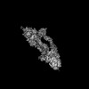 39184MC  8ydpC  8ydqC  8ydrC  8ydsC  8ydtC  8yduC  8ydvC  8ydwC  8ydyC  8ydzC M: map data used to model this data C: citing same article ( |
|---|---|
| Similar structure data |

-Links
 PDBj
PDBj


- Assembly
| Deposited unit | 
|
|---|---|
| 1 |
|
-Components
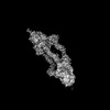
| #1: Protein/peptide | Mass: 4721.475 Da / Num. of mol.: 3 / Source method: obtained synthetically / Source: (synth.) synthetic construct (others) #2: Protein | Mass: 144226.750 Da / Num. of mol.: 3 / Mutation: F817P, A892P, A898P, A942P, K986P, V987P Source method: isolated from a genetically manipulated source Details: the N-terminal sequence (from -18 to 12) is an mammalian secretion signal sequence, and the C-terminal sequence includes "T4 fibritin trimerization mottif" (Gly1211-Leu1237), "HRV 3C ...Details: the N-terminal sequence (from -18 to 12) is an mammalian secretion signal sequence, and the C-terminal sequence includes "T4 fibritin trimerization mottif" (Gly1211-Leu1237), "HRV 3C protease cleavage site" (Leu1241-Pro1248), "8x His tag" (His1250-His1257) and "Twin-strep tag" (Trp1260-Lys1288) Source: (gene. exp.) Severe acute respiratory syndrome coronavirus 2Gene: S, 2 / Cell line (production host): Expi293F / Production host:  Homo sapiens (human) / References: UniProt: P0DTC2 Homo sapiens (human) / References: UniProt: P0DTC2#3: Sugar | ChemComp-NAG /   Type: D-saccharide, beta linking / Mass: 221.208 Da / Num. of mol.: 27 / Source method: obtained synthetically / Formula: C8H15NO6 Type: D-saccharide, beta linking / Mass: 221.208 Da / Num. of mol.: 27 / Source method: obtained synthetically / Formula: C8H15NO6Has ligand of interest | N | Has protein modification | Y | |
|---|
-Experimental details
-Experiment
| Experiment | Method: ELECTRON MICROSCOPY |
|---|---|
| EM experiment | Aggregation state: PARTICLE / 3D reconstruction method: single particle reconstruction |
- Sample preparation
| Component | Name: SARS-CoV-2 spike ectodomain (HexaPro, Omicron BA.2 variant) in complex with CeSPIACE Type: COMPLEX / Entity ID: #1-#2 / Source: RECOMBINANT | ||||||||||||||||
|---|---|---|---|---|---|---|---|---|---|---|---|---|---|---|---|---|---|
| Molecular weight | Experimental value: NO | ||||||||||||||||
| Source (natural) | Organism: Severe acute respiratory syndrome coronavirus 2 | ||||||||||||||||
| Source (recombinant) | Organism: Homo sapiens (human) | ||||||||||||||||
| Buffer solution | pH: 8 | ||||||||||||||||
| Buffer component |
| ||||||||||||||||
| Specimen | Conc.: 0.42 mg/ml / Embedding applied: NO / Shadowing applied: NO / Staining applied: NO / Vitrification applied: YES | ||||||||||||||||
| Specimen support | Grid material: GOLD / Grid mesh size: 300 divisions/in. / Grid type: Quantifoil R1.2/1.3 | ||||||||||||||||
| Vitrification | Instrument: FEI VITROBOT MARK IV / Cryogen name: ETHANE / Humidity: 95 % / Chamber temperature: 278 K |
- Electron microscopy imaging
| Microscopy | Model: JEOL CRYO ARM 300 |
|---|---|
| Electron gun | Electron source:  FIELD EMISSION GUN / Accelerating voltage: 300 kV / Illumination mode: FLOOD BEAM FIELD EMISSION GUN / Accelerating voltage: 300 kV / Illumination mode: FLOOD BEAM |
| Electron lens | Mode: BRIGHT FIELD / Nominal defocus max: 2500 nm / Nominal defocus min: 1500 nm / Cs: 3.4 mm / C2 aperture diameter: 50 µm / Alignment procedure: COMA FREE |
| Specimen holder | Cryogen: NITROGEN / Specimen holder model: JEOL 3200FSC CRYOHOLDER |
| Image recording | Electron dose: 69.6 e/Å2 / Detector mode: COUNTING / Film or detector model: GATAN K2 SUMMIT (4k x 4k) |
- Processing
| EM software |
| ||||||||||||||||||||||||||||||||||||||||
|---|---|---|---|---|---|---|---|---|---|---|---|---|---|---|---|---|---|---|---|---|---|---|---|---|---|---|---|---|---|---|---|---|---|---|---|---|---|---|---|---|---|
| CTF correction | Type: PHASE FLIPPING AND AMPLITUDE CORRECTION | ||||||||||||||||||||||||||||||||||||||||
| Symmetry | Point symmetry: C1 (asymmetric) | ||||||||||||||||||||||||||||||||||||||||
| 3D reconstruction | Resolution: 4.9 Å / Resolution method: FSC 0.143 CUT-OFF / Num. of particles: 111423 / Symmetry type: POINT | ||||||||||||||||||||||||||||||||||||||||
| Atomic model building | Protocol: RIGID BODY FIT | ||||||||||||||||||||||||||||||||||||||||
| Atomic model building | PDB-ID: 6XKL Accession code: 6XKL / Source name: PDB / Type: experimental model |