 Movie
Movie Controller

[English] 日本語
 Yorodumi
- PDB-8yai: Crystal structure of glucose 1-dehydrogenase mutant1 from Limosil... -
+ Open data

- Basic information
| Entry | Database: PDB / ID: 8yai | ||||||
|---|---|---|---|---|---|---|---|
| Title | Crystal structure of glucose 1-dehydrogenase mutant1 from Limosilactobacillus fermentum | ||||||
 Components Components | SDR family oxidoreductase | ||||||
 Keywords Keywords | HYDROLASE / glucose 1-dehydrogenase | ||||||
| Function / homology | PKS_KR / Short-chain dehydrogenase/reductase, conserved site / Short-chain dehydrogenases/reductases family signature. / Enoyl-(Acyl carrier protein) reductase / Short-chain dehydrogenase/reductase SDR / oxidoreductase activity / NAD(P)-binding domain superfamily / SDR family oxidoreductase Function and homology information Function and homology information | ||||||
| Biological species |  Limosilactobacillus fermentum (bacteria) Limosilactobacillus fermentum (bacteria) | ||||||
| Method |  X-RAY DIFFRACTION / SYNCHROTRON / MOLECULAR REPLACEMENT / Resolution: 2.13 Å X-RAY DIFFRACTION / SYNCHROTRON / MOLECULAR REPLACEMENT / Resolution: 2.13 Å | ||||||
 Authors Authors | Cong, L. / Wang, J.J. / Wei, H.L. / Liu, W.D. / You, S. | ||||||
| Funding support |  China, 1items China, 1items
| ||||||
 Citation Citation | Journal: Adv.Synth.Catal. / Year: 2024 Title: Structure-Guided Engineering of a Short-Chain Dehydrogenase LfSDR1 for Efficient Biosynthesis of (R)-9-(2-Hydroxypropyl)adenine, the Key Intermediate of Tenofovir Authors: Wang, Q. / Cong, L. / Guo, J. / Wang, J. / Han, X. / Zhang, W. / Liu, W. / Wei, H. / You, S. | ||||||
| History |
|
- Structure visualization
| Structure viewer | Molecule: MolmilJmol/JSmol |
|---|
- Downloads & links
-Download
| PDBx/mmCIF format | 8yai.cif.gz | 184.6 KB | Display | PDBx/mmCIF format |
|---|---|---|---|---|
| PDB format | pdb8yai.ent.gz | 146.1 KB | Display | PDB format |
| PDBx/mmJSON format | 8yai.json.gz | Tree view | PDBx/mmJSON format | |
| Others |  Other downloads Other downloads |
-Validation report
| Summary document | 8yai_validation.pdf.gz | 458.3 KB | Display | wwPDB validaton report |
|---|---|---|---|---|
| Full document | 8yai_full_validation.pdf.gz | 468.9 KB | Display | |
| Data in XML | 8yai_validation.xml.gz | 41.8 KB | Display | |
| Data in CIF | 8yai_validation.cif.gz | 55 KB | Display | |
| Arichive directory | https://data.pdbj.org/pub/pdb/validation_reports/ya/8yaiftp://data.pdbj.org/pub/pdb/validation_reports/ya/8yai | HTTPS FTP |
-Related structure data
| Related structure data |  8yauC  8zaxC 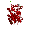 7p7yS S: Starting model for refinement C: citing same article ( |
|---|---|
| Similar structure data |
-Links
 PDBj
PDBj


- Assembly
| Deposited unit | 
| |||||||||||||||||||||||||||||||||||||||||||||||||||||||||||||||||||||||||||||||||||||||||||||||
|---|---|---|---|---|---|---|---|---|---|---|---|---|---|---|---|---|---|---|---|---|---|---|---|---|---|---|---|---|---|---|---|---|---|---|---|---|---|---|---|---|---|---|---|---|---|---|---|---|---|---|---|---|---|---|---|---|---|---|---|---|---|---|---|---|---|---|---|---|---|---|---|---|---|---|---|---|---|---|---|---|---|---|---|---|---|---|---|---|---|---|---|---|---|---|---|---|
| 1 | 
| |||||||||||||||||||||||||||||||||||||||||||||||||||||||||||||||||||||||||||||||||||||||||||||||
| Unit cell |
| |||||||||||||||||||||||||||||||||||||||||||||||||||||||||||||||||||||||||||||||||||||||||||||||
| Noncrystallographic symmetry (NCS) | NCS domain:
NCS domain segments: Ens-ID: 1
|