 Movie
Movie Controller


+ Open data

- Basic information
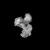
| Entry | Database: PDB / ID: 8yah | |||||||||||||||||||||||||||||||||||||||||||||
|---|---|---|---|---|---|---|---|---|---|---|---|---|---|---|---|---|---|---|---|---|---|---|---|---|---|---|---|---|---|---|---|---|---|---|---|---|---|---|---|---|---|---|---|---|---|---|
| Title | full length AP5 complex bound to SPG11-SPG15 | |||||||||||||||||||||||||||||||||||||||||||||
 Components Components |
| |||||||||||||||||||||||||||||||||||||||||||||
 Keywords Keywords | TRANSPORT PROTEIN / Complex | |||||||||||||||||||||||||||||||||||||||||||||
| Function / homology |  Function and homology information Function and homology informationAP-5 adaptor complex / phagosome-lysosome fusion involved in apoptotic cell clearance / late endosome to Golgi transport / walking behavior / AP-type membrane coat adaptor complex / corticospinal tract morphogenesis / regulation of store-operated calcium entry / localization within membrane / autophagosome organization / lysosomal protein catabolic process ...AP-5 adaptor complex / phagosome-lysosome fusion involved in apoptotic cell clearance / late endosome to Golgi transport / walking behavior / AP-type membrane coat adaptor complex / corticospinal tract morphogenesis / regulation of store-operated calcium entry / localization within membrane / autophagosome organization / lysosomal protein catabolic process / axon extension / motor neuron apoptotic process / vesicle transport along microtubule / axo-dendritic transport / cholesterol efflux / lysosome organization / endosomal transport / axon development / Golgi organization / motor behavior / synaptic vesicle transport / neuromuscular junction development / autophagosome assembly / skeletal muscle fiber development / vesicle-mediated transport / axonogenesis / protein catabolic process / intracellular protein transport / protein import into nucleus / double-strand break repair via homologous recombination / autophagy / memory / late endosome / late endosome membrane / protein transport / gene expression / cytoplasmic vesicle / chemical synaptic transmission / lysosome / nuclear speck / axon / lysosomal membrane / dendrite / synapse / protein kinase binding / nucleolus / endoplasmic reticulum / nucleoplasm / membrane / nucleus / plasma membrane / cytosol / cytoplasm Similarity search - Function | |||||||||||||||||||||||||||||||||||||||||||||
| Biological species |   Homo sapiens (human) Homo sapiens (human) | |||||||||||||||||||||||||||||||||||||||||||||
| Method | ELECTRON MICROSCOPY / single particle reconstruction / cryo EM / Resolution: 3.3 Å | |||||||||||||||||||||||||||||||||||||||||||||
 Authors Authors | Su, M.-Y. | |||||||||||||||||||||||||||||||||||||||||||||
| Funding support |  China, 1items China, 1items
| |||||||||||||||||||||||||||||||||||||||||||||
 Citation Citation | Journal: Nat Struct Mol Biol / Year: 2025 Title: Structural basis for membrane remodeling by the AP5-SPG11-SPG15 complex. Authors: Xinyi Mai / Yang Wang / Xi Wang / Ming Liu / Fei Teng / Zheng Liu / Ming-Yuan Su / Goran Stjepanovic / Abstract: The human spastizin (spastic paraplegia 15, SPG15) and spatacsin (spastic paraplegia 11, SPG11) complex is involved in the formation of lysosomes, and mutations in these two proteins are linked with ...The human spastizin (spastic paraplegia 15, SPG15) and spatacsin (spastic paraplegia 11, SPG11) complex is involved in the formation of lysosomes, and mutations in these two proteins are linked with hereditary autosomal-recessive spastic paraplegia. SPG11-SPG15 can cooperate with the fifth adaptor protein complex (AP5) involved in membrane sorting of late endosomes. We employed cryogenic-electron microscopy and in silico predictions to investigate the structural assemblies of the SPG11-SPG15 and AP5-SPG11-SPG15 complexes. The W-shaped SPG11-SPG15 intertwined in a head-to-head fashion, and the N-terminal region of SPG11 is required for AP5 complex interaction and assembly. The AP5 complex is in a super-open conformation. Our findings reveal that the AP5-SPG11-SPG15 complex can bind PI3P molecules, sense membrane curvature and drive membrane remodeling in vitro. These studies provide insights into the structure and function of the spastic paraplegia AP5-SPG11-SPG15 complex, which is essential for the initiation of autolysosome tubulation. | |||||||||||||||||||||||||||||||||||||||||||||
| History |
|
- Structure visualization
| Structure viewer | Molecule: MolmilJmol/JSmol |
|---|
- Downloads & links
-Download
| PDBx/mmCIF format | 8yah.cif.gz | 445.6 KB | Display | PDBx/mmCIF format |
|---|---|---|---|---|
| PDB format | pdb8yah.ent.gz | 308.4 KB | Display | PDB format |
| PDBx/mmJSON format | 8yah.json.gz | Tree view | PDBx/mmJSON format | |
| Others |  Other downloads Other downloads |
-Validation report
| Arichive directory | https://data.pdbj.org/pub/pdb/validation_reports/ya/8yahftp://data.pdbj.org/pub/pdb/validation_reports/ya/8yah | HTTPS FTP |
|---|
-Related structure data
| Related structure data | 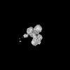 39099MC  8yabC  8yadC C: citing same article ( M: map data used to model this data |
|---|---|
| Similar structure data |
-Links
 PDBj
PDBj
- Assembly
| Deposited unit | 
|
|---|---|
| 1 |
|
-Components
| #1: Protein | Mass: 89574.422 Da / Num. of mol.: 1 Source method: isolated from a genetically manipulated source Source: (gene. exp.) Homo sapiens (human) / References: UniProt: Q3U829 |
|---|---|
| #2: Protein | Mass: 94038.109 Da / Num. of mol.: 1 Source method: isolated from a genetically manipulated source Source: (gene. exp.) Homo sapiens (human) / Gene: AP5B1 / Cell line (production host): HEK293 / Production host: Homo sapiens (human) / References: UniProt: Q2VPB7 |
| #3: Protein | Mass: 22550.984 Da / Num. of mol.: 1 Source method: isolated from a genetically manipulated source Source: (gene. exp.) Homo sapiens (human) / Gene: AP5S1 / Cell line (production host): HEK293 / Production host: Homo sapiens (human) / References: UniProt: Q9NUS5 |
| #4: Protein | Mass: 54393.285 Da / Num. of mol.: 1 Source method: isolated from a genetically manipulated source Source: (gene. exp.) Homo sapiens (human) / References: UniProt: Q8BJ63 |
| #5: Protein | Mass: 279182.594 Da / Num. of mol.: 1 Source method: isolated from a genetically manipulated source Source: (gene. exp.) Homo sapiens (human) / Gene: SPG11 / Cell line (production host): HEK293 / Production host: Homo sapiens (human) / References: UniProt: Q96JI7 |
| Has protein modification | N |
-Experimental details
-Experiment
| Experiment | Method: ELECTRON MICROSCOPY |
|---|---|
| EM experiment | Aggregation state: PARTICLE / 3D reconstruction method: single particle reconstruction |
- Sample preparation
| Component | Name: full length AP5 complex bound to SPG11-SPG15 / Type: COMPLEX / Entity ID: all / Source: RECOMBINANT |
|---|---|
| Molecular weight | Value: 0.826 MDa / Experimental value: NO |
| Source (natural) | Organism: Homo sapiens (human) |
| Source (recombinant) | Organism: Homo sapiens (human) |
| Buffer solution | pH: 7.4 |
| Specimen | Embedding applied: NO / Shadowing applied: NO / Staining applied: NO / Vitrification applied: YES |
| Vitrification | Cryogen name: ETHANE |
- Electron microscopy imaging
| Experimental equipment |  Model: Titan Krios / Image courtesy: FEI Company |
|---|---|
| Microscopy | Model: FEI TITAN KRIOS |
| Electron gun | Electron source:  FIELD EMISSION GUN / Accelerating voltage: 300 kV / Illumination mode: SPOT SCAN FIELD EMISSION GUN / Accelerating voltage: 300 kV / Illumination mode: SPOT SCAN |
| Electron lens | Mode: BRIGHT FIELD / Nominal defocus max: 1800 nm / Nominal defocus min: 1200 nm |
| Image recording | Electron dose: 1.2386 e/Å2 / Film or detector model: GATAN K3 (6k x 4k) |
- Processing
| EM software | Name: PHENIX / Category: model refinement | ||||||||||||||||||||||||
|---|---|---|---|---|---|---|---|---|---|---|---|---|---|---|---|---|---|---|---|---|---|---|---|---|---|
| CTF correction | Type: NONE | ||||||||||||||||||||||||
| 3D reconstruction | Resolution: 3.3 Å / Resolution method: FSC 0.143 CUT-OFF / Num. of particles: 586806 / Symmetry type: POINT | ||||||||||||||||||||||||
| Refine LS restraints |
|
