 Movie
Movie Controller


+ Open data

- Basic information
| Entry | 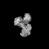 | |||||||||
|---|---|---|---|---|---|---|---|---|---|---|
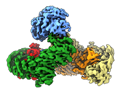
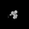
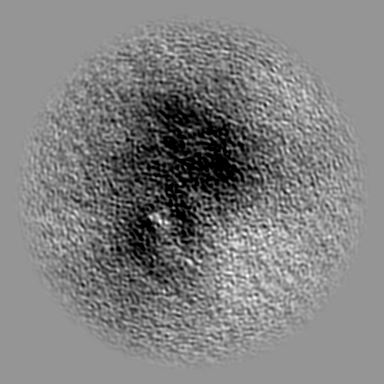
| Title | AP5 complex bound to SPG11-SPG15 | |||||||||
 Map data Map data | ||||||||||
 Sample Sample |
| |||||||||
 Keywords Keywords | Complex / TRANSPORT PROTEIN | |||||||||
| Function / homology |  Function and homology information Function and homology informationAP-5 adaptor complex / phagosome-lysosome fusion involved in apoptotic cell clearance / late endosome to Golgi transport / walking behavior / AP-type membrane coat adaptor complex / corticospinal tract morphogenesis / autophagosome organization / regulation of store-operated calcium entry / lysosomal protein catabolic process / motor neuron apoptotic process ...AP-5 adaptor complex / phagosome-lysosome fusion involved in apoptotic cell clearance / late endosome to Golgi transport / walking behavior / AP-type membrane coat adaptor complex / corticospinal tract morphogenesis / autophagosome organization / regulation of store-operated calcium entry / lysosomal protein catabolic process / motor neuron apoptotic process / localization within membrane / axon extension / motor behavior / vesicle transport along microtubule / axo-dendritic transport / cholesterol efflux / lysosome organization / endosomal transport / axon development / Golgi organization / synaptic vesicle transport / neuromuscular junction development / autophagosome assembly / skeletal muscle fiber development / axonogenesis / vesicle-mediated transport / protein catabolic process / protein import into nucleus / intracellular protein transport / memory / double-strand break repair via homologous recombination / autophagy / gene expression / late endosome / late endosome membrane / protein transport / cytoplasmic vesicle / chemical synaptic transmission / lysosome / nuclear speck / axon / lysosomal membrane / synapse / dendrite / nucleolus / protein kinase binding / endoplasmic reticulum / nucleoplasm / membrane / nucleus / plasma membrane / cytosol / cytoplasm Similarity search - Function | |||||||||
| Biological species |  Homo sapiens (human) / Homo sapiens (human) /  | |||||||||
| Method | single particle reconstruction / cryo EM / Resolution: 3.26 Å | |||||||||
 Authors Authors | Su M-Y | |||||||||
| Funding support |  China, 1 items China, 1 items
| |||||||||
 Citation Citation | Journal: Nat Struct Mol Biol / Year: 2025 Title: Structural basis for membrane remodeling by the AP5-SPG11-SPG15 complex. Authors: Xinyi Mai / Yang Wang / Xi Wang / Ming Liu / Fei Teng / Zheng Liu / Ming-Yuan Su / Goran Stjepanovic / Abstract: The human spastizin (spastic paraplegia 15, SPG15) and spatacsin (spastic paraplegia 11, SPG11) complex is involved in the formation of lysosomes, and mutations in these two proteins are linked with ...The human spastizin (spastic paraplegia 15, SPG15) and spatacsin (spastic paraplegia 11, SPG11) complex is involved in the formation of lysosomes, and mutations in these two proteins are linked with hereditary autosomal-recessive spastic paraplegia. SPG11-SPG15 can cooperate with the fifth adaptor protein complex (AP5) involved in membrane sorting of late endosomes. We employed cryogenic-electron microscopy and in silico predictions to investigate the structural assemblies of the SPG11-SPG15 and AP5-SPG11-SPG15 complexes. The W-shaped SPG11-SPG15 intertwined in a head-to-head fashion, and the N-terminal region of SPG11 is required for AP5 complex interaction and assembly. The AP5 complex is in a super-open conformation. Our findings reveal that the AP5-SPG11-SPG15 complex can bind PI3P molecules, sense membrane curvature and drive membrane remodeling in vitro. These studies provide insights into the structure and function of the spastic paraplegia AP5-SPG11-SPG15 complex, which is essential for the initiation of autolysosome tubulation. | |||||||||
| History |
|
- Structure visualization
| Supplemental images |
|---|
- Downloads & links
-EMDB archive
| Map data | emd_39094.map.gz | 107.2 MB | EMDB map data format | |
|---|---|---|---|---|
| Header (meta data) | emd-39094-v30.xmlemd-39094.xml | 27.7 KB 27.7 KB | Display Display | EMDB header |
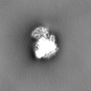
| Images |  emd_39094.png emd_39094.png | 115 KB | ||
| Filedesc metadata | emd-39094.cif.gz | 7.8 KB | ||
| Others | emd_39094_additional_1.map.gzemd_39094_additional_2.map.gzemd_39094_half_map_1.map.gzemd_39094_half_map_2.map.gz | 184.6 MB 191.2 MB 200.7 MB 200.7 MB | ||
| Archive directory |  http://ftp.pdbj.org/pub/emdb/structures/EMD-39094ftp://ftp.pdbj.org/pub/emdb/structures/EMD-39094 http://ftp.pdbj.org/pub/emdb/structures/EMD-39094ftp://ftp.pdbj.org/pub/emdb/structures/EMD-39094 | HTTPS FTP |
-Related structure data
| Related structure data |  8yabMC  8yadC  8yahC M: atomic model generated by this map C: citing same article ( |
|---|---|
| Similar structure data |
-Links
| EMDB pages | EMDB (EBI/PDBe) / EMDataResource |
|---|---|
| Related items in Molecule of the Month |

-Map
| File | Download / File: emd_39094.map.gz / Format: CCP4 / Size: 216 MB / Type: IMAGE STORED AS FLOATING POINT NUMBER (4 BYTES) | ||||||||||||||||||||||||||||||||||||
|---|---|---|---|---|---|---|---|---|---|---|---|---|---|---|---|---|---|---|---|---|---|---|---|---|---|---|---|---|---|---|---|---|---|---|---|---|---|
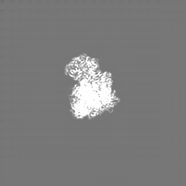
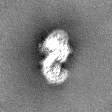
| Projections & slices | Image control
Images are generated by Spider. | ||||||||||||||||||||||||||||||||||||
| Voxel size | X=Y=Z: 0.85 Å | ||||||||||||||||||||||||||||||||||||
| Density |
| ||||||||||||||||||||||||||||||||||||
| Symmetry | Space group: 1 | ||||||||||||||||||||||||||||||||||||
| Details | EMDB XML:
|
 Z (Sec.)
 Y (Row.)
 X (Col.)


-Supplemental data
-Additional map: deepEMhancer map highRes model
| File | emd_39094_additional_1.map | ||||||||||||
|---|---|---|---|---|---|---|---|---|---|---|---|---|---|
| Annotation | deepEMhancer map highRes model | ||||||||||||
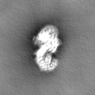
| Projections & Slices |
| ||||||||||||
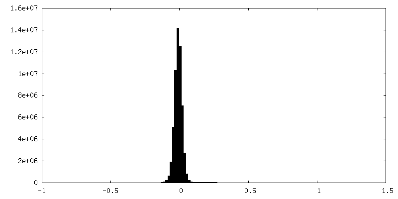
| Density Histograms |


-Additional map: deepEMhancer map tightTarget model
| File | emd_39094_additional_2.map | ||||||||||||
|---|---|---|---|---|---|---|---|---|---|---|---|---|---|
| Annotation | deepEMhancer map tightTarget model | ||||||||||||
| Projections & Slices |
| ||||||||||||
| Density Histograms |


-Half map: #1
| File | emd_39094_half_map_1.map | ||||||||||||
|---|---|---|---|---|---|---|---|---|---|---|---|---|---|
| Projections & Slices |
| ||||||||||||
| Density Histograms |


-Half map: #2
| File | emd_39094_half_map_2.map | ||||||||||||
|---|---|---|---|---|---|---|---|---|---|---|---|---|---|
| Projections & Slices |
| ||||||||||||
| Density Histograms |


- Sample components
-Entire : AP5 complex bound to SPG11-SPG15
| Entire | Name: AP5 complex bound to SPG11-SPG15 |
|---|---|
| Components |
|
-Supramolecule #1: AP5 complex bound to SPG11-SPG15
| Supramolecule | Name: AP5 complex bound to SPG11-SPG15 / type: complex / ID: 1 / Parent: 0 / Macromolecule list: all |
|---|---|
| Source (natural) | Organism: Homo sapiens (human) |
| Molecular weight | Theoretical: 600 KDa |
-Macromolecule #1: AP-5 complex subunit zeta-1
| Macromolecule | Name: AP-5 complex subunit zeta-1 / type: protein_or_peptide / ID: 1 / Number of copies: 1 / Enantiomer: LEVO |
|---|---|
| Source (natural) | Organism: |
| Molecular weight | Theoretical: 89.574422 KDa |
| Recombinant expression | Organism: Homo sapiens (human) |
| Sequence | String: MAFSAGAESL LHQAREIQDE ELRRFCSRVT KLLQEAPGPA TVDALQRLFL IVSATKYPRR LEKMCVDLLQ TTLCLPASPE QLQVLCAAI LREMSPFNDL ALSCDHTPNT RQLSLVASVL LAQGDRKGEI RCVSQRIFKI LENRQPEGPS VRPLLPILSK V IGLAPGIL ...String: MAFSAGAESL LHQAREIQDE ELRRFCSRVT KLLQEAPGPA TVDALQRLFL IVSATKYPRR LEKMCVDLLQ TTLCLPASPE QLQVLCAAI LREMSPFNDL ALSCDHTPNT RQLSLVASVL LAQGDRKGEI RCVSQRIFKI LENRQPEGPS VRPLLPILSK V IGLAPGIL MEDQTNLLSK RLVDWLRYAS IQQGLPYSGG FFSTPRTRQP GPITEVDGAV ASDFFTVLST GQHFTEDQWV NM QAFSMLR KWLLHSGPED PCSPDADDKS ELEGSTLSVL SAASTASRLL PPRERLREVA FEYCQRLLEQ SNRRALRKGD SDL QKACLV EAVSVLDVLC RQDPSFLYRT LSCLKALHRR LGEDPGSERA LVPLAQFFLN HGEAAAMDAE AVYGQLLRGL PSER FHSPT LAFEVIHFCT HNLALFDSHF LSLLRLSFPS LFKFLAWNSP PLTAEFVVLL PALVDAGTAV EMLHALLDLP CLTAA LDLQ LRSTQTPSER LLWDISLRVP SCLEAFQDPQ FQGLFRHLLR TKASGSTERL TPLHQVLKPM ASCARVTQCA EAVPVL LQA FFSAVTQTAD GALINQLALL LLERSDSLYP VPQYEARVHG VLSSQLLVLC KLKPSLVVEL SRELLEFVGS VSSIHSR AS VFTCVVWAIG EYLSVTWDKR CTAEQINKFF EALEALLFEV TQSRPLADLP CCPPEVVTAL MTTLTKLASR SQDLIPRV S LFLSKMRTLA QNPATSSVHS EEGAESIRTR ASELLTLLKM PSVAQFVFTP PAGVCQPRYH RDTNVALPLA LRTVSRLVE KEAGLLPG UniProtKB: AP-5 complex subunit zeta-1 |
-Macromolecule #2: AP-5 complex subunit beta-1
| Macromolecule | Name: AP-5 complex subunit beta-1 / type: protein_or_peptide / ID: 2 / Number of copies: 1 / Enantiomer: LEVO |
|---|---|
| Source (natural) | Organism: Homo sapiens (human) |
| Molecular weight | Theoretical: 94.038109 KDa |
| Recombinant expression | Organism: Homo sapiens (human) |
| Sequence | String: MGPLSRDAWA QRLGAFRASP SAFMAGPEGE DLGRDLLSDL RSEKLSEQTK VSLLALSMEY PAQLWPDASA AEVAATSLLD TLVLLPPRP SALRRPLLLA ATTALAAGGA LGPTSGASCR LLPLLLGLAA GSDLGRGFVP ASEQRPLQAT ACECLRELES C KPGLLGGS ...String: MGPLSRDAWA QRLGAFRASP SAFMAGPEGE DLGRDLLSDL RSEKLSEQTK VSLLALSMEY PAQLWPDASA AEVAATSLLD TLVLLPPRP SALRRPLLLA ATTALAAGGA LGPTSGASCR LLPLLLGLAA GSDLGRGFVP ASEQRPLQAT ACECLRELES C KPGLLGGS LGLLRGLLGQ EGPVQPLSLL LALALRNTLV LQSRVGAGLG GLLTDKVSPT GGGPWDWTLV EEGDGRLQPQ AP SWPAAEE GEGERSLTAR EHSPEEAREL RAAVIQLLDT SYLLTPVAQA QLLWLLGWAL RGLQGQPPAL FKPQLVRLLG TAQ LTLLHA MLALKAAFGE ALFTAQDEAL LLRRLTLAAQ HPALPPPTHL FYLHCVLSFP ENWPLGPEGE EAAPLLLGPQ LCRG LLPSL LHDPMALLAR LHLLCLLCAE EEEEEKGQLP SPRHYLEELL AGLRQRAALD GGPRALATLC FQASYLVACC LAGQP TVLT PLIHGLAQLY QARPMLAPHF VDLLDQVDSE LREPLKVVLR QVVVSRPGRD EALCWHLQML AKVADGDAQS ATLNFL QAA AAHCTNWDLQ QGLLRVCRAL LRAGVRGGLV DLLQVLARQL EDPDGRDHAR LYYILLAHLA APKLGVALGP SLAAPAL AS SLVAENQGFV AALMVQEAPA LVRLSLGSHR VKGPLPVLKL QPEALEPIYS LELRFRVEGQ LYAPLEAVHV PCLCPGRP A RPLLLPLQPR CPAPARLDVH ALYTTSTGLT CHAHLPPLFV NFADLFLPFP QPPEGAGLGF FEELWDSCLP EGAESRVWC PLGPQGLEGL VSRHLEPFVV VAQPPTSYCV AIHLPPDSKL LLRLEAALAD GVPVALRTDD WAVLPLAGDY LRGLAAAV UniProtKB: AP-5 complex subunit beta-1 |
-Macromolecule #3: AP-5 complex subunit sigma-1
| Macromolecule | Name: AP-5 complex subunit sigma-1 / type: protein_or_peptide / ID: 3 / Number of copies: 1 / Enantiomer: LEVO |
|---|---|
| Source (natural) | Organism: Homo sapiens (human) |
| Molecular weight | Theoretical: 22.550984 KDa |
| Recombinant expression | Organism: Homo sapiens (human) |
| Sequence | String: MVHAFLIHTL RAPNTEDTGL CRVLYSCVFG AEKSPDDPRP HGAERDRLLR KEQILAVARQ VESMCRLQQQ ASGRPPMDLQ PQSSDEQVP LHEAPRGAFR LAAENPFQEP RTVVWLGVLS LGFALVLDAH ENLLLAEGTL RLLTRLLLDH LRLLAPSTSL L LRADRIEG ...String: MVHAFLIHTL RAPNTEDTGL CRVLYSCVFG AEKSPDDPRP HGAERDRLLR KEQILAVARQ VESMCRLQQQ ASGRPPMDLQ PQSSDEQVP LHEAPRGAFR LAAENPFQEP RTVVWLGVLS LGFALVLDAH ENLLLAEGTL RLLTRLLLDH LRLLAPSTSL L LRADRIEG ILTRFLPHGQ LLFLNDQFVQ GLEKEFSAAW PR UniProtKB: AP-5 complex subunit sigma-1 |
-Macromolecule #4: AP-5 complex subunit mu-1
| Macromolecule | Name: AP-5 complex subunit mu-1 / type: protein_or_peptide / ID: 4 / Number of copies: 1 / Enantiomer: LEVO |
|---|---|
| Source (natural) | Organism: |
| Molecular weight | Theoretical: 54.393285 KDa |
| Recombinant expression | Organism: Homo sapiens (human) |
| Sequence | String: MALRAVWLIR HEPGTPLGGT VRFSRRYPTV EKRAKAFNGM TYVPVPEDGP FLRALLFQLR LLDDDKDFME RRDGCSRINK TSIYGLSVG GEELWPVIAF LRDSMIYASV PLVEQALSPR PPLISISGVS QGLELLLGIQ DFLYSSQKND TDLHTKLSQL P DLLLQACP ...String: MALRAVWLIR HEPGTPLGGT VRFSRRYPTV EKRAKAFNGM TYVPVPEDGP FLRALLFQLR LLDDDKDFME RRDGCSRINK TSIYGLSVG GEELWPVIAF LRDSMIYASV PLVEQALSPR PPLISISGVS QGLELLLGIQ DFLYSSQKND TDLHTKLSQL P DLLLQACP LGTLLDANLQ NSLNSINSVS VTQPQKQPAW KVGAYKGKAQ ISISITETVK CMQYGKQDIA DTWQVAGTVA CK CDLEGVM PAVTISLSLP TNGSPLQDII VHPCVTSLDS AILTSSSIDT MDDSAFSGPY KFPFTPPLES FNLCHYTSQV PVP PILGSY HMKEEGVQLK VTVNFKLHES VRNNFEVCEA HIPFYNRGPI THLEYKASFG QLEVFREKSL LVWIIGQKFP KSME ISLSG TLTFGVKGHN KQPFDHICIG NTAYIKLNFR IADYTLTGCY ADQHSVQVFA SGKPKISAYR KLISSDYYIW NSKAP APVT YASLLP UniProtKB: AP-5 complex subunit mu-1 |
-Macromolecule #5: Spatacsin
| Macromolecule | Name: Spatacsin / type: protein_or_peptide / ID: 5 / Number of copies: 1 / Enantiomer: LEVO |
|---|---|
| Source (natural) | Organism: Homo sapiens (human) |
| Molecular weight | Theoretical: 58.115113 KDa |
| Recombinant expression | Organism: Homo sapiens (human) |
| Sequence | String: MAAEEGVASA ASAGGSWGTA AMGRVLPMLL VPVPAEAMGQ LGSRAQLRTQ PEALGSLTAA GSLQVLSLTP GSRGGGRCCL EGPFWHFLW EDSRNSSTPT EKPKLLALGE NYELLIYEFN LKDGRCDATI LYSCSREALQ KLIDDQDISI SLLSLRILSF H NNTSLLFI ...String: MAAEEGVASA ASAGGSWGTA AMGRVLPMLL VPVPAEAMGQ LGSRAQLRTQ PEALGSLTAA GSLQVLSLTP GSRGGGRCCL EGPFWHFLW EDSRNSSTPT EKPKLLALGE NYELLIYEFN LKDGRCDATI LYSCSREALQ KLIDDQDISI SLLSLRILSF H NNTSLLFI NKCVILHIIF PERDAAIRVL NCFTLPLPAQ AVDMIIDTQL CRGILFVLSS LGWIYIFDVV DGTYVAHVDL AL HKEDMCN EQQQEPAKIS SFTSLKVSQD LDVAVIVSSS NSAVALNLNL YFRQHPGHLL CERILEDLPI QGPKGVDEDD PVN SAYNMK LAKFSFQIDR SWKAQLSSLN ETIKNSKLEV SCCAPWFQDI LHLESPESGN HSTSVQSWAF IPQDIMHGQY NVLQ KDHAK TSDPGRSWKI MHISEQEEPI ELKCVSVTGF TALFTWEVER MGYTITLWDL ETQGMQCFSL GTKCIPVDSS GDQQL CFVL TENGLSLILF GLTQEEFLNR LMIHGSASTV DTLCHLNGWG R UniProtKB: Spatacsin |
-Experimental details
-Structure determination
| Method | cryo EM |
|---|---|
 Processing Processing | single particle reconstruction |
| Aggregation state | particle |
-Sample preparation
| Buffer | pH: 7.4 |
|---|---|
| Vitrification | Cryogen name: ETHANE |
- Electron microscopy
| Microscope | FEI TITAN KRIOS |
|---|---|
| Image recording | Film or detector model: GATAN K3 (6k x 4k) / Average electron dose: 1.2386 e/Å2 |
| Electron beam | Acceleration voltage: 300 kV / Electron source:  FIELD EMISSION GUN FIELD EMISSION GUN |
| Electron optics | Illumination mode: SPOT SCAN / Imaging mode: BRIGHT FIELD / Nominal defocus max: 1.8 µm / Nominal defocus min: 1.2 µm |
| Experimental equipment |  Model: Titan Krios / Image courtesy: FEI Company |
