 Movie
Movie Controller

[English] 日本語
 Yorodumi
- PDB-8y6z: Crystal structure of the Marinitoga sp. Csx1-Crn2 fusion ribonucl... -
+ Open data

- Basic information
| Entry | Database: PDB / ID: 8y6z | ||||||
|---|---|---|---|---|---|---|---|
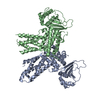
| Title | Crystal structure of the Marinitoga sp. Csx1-Crn2 fusion ribonuclease of type III CRISPR | ||||||
 Components Components | CRISPR-associated protein | ||||||
 Keywords Keywords | HYDROLASE / type III CRISPR / self-limiting ribonuclease / ring nuclease / Csx1 / Crn2. | ||||||
| Function / homology | STIV B116-like / STIV B116-like superfamily / STIV B116-like / CRISPR-associated protein DxTHG, conserved site / CRISPR-associated protein Function and homology information Function and homology information | ||||||
| Biological species |  Marinitoga sp. 1155 (bacteria) Marinitoga sp. 1155 (bacteria) | ||||||
| Method |  X-RAY DIFFRACTION / SYNCHROTRON / MOLECULAR REPLACEMENT / Resolution: 3.65 Å X-RAY DIFFRACTION / SYNCHROTRON / MOLECULAR REPLACEMENT / Resolution: 3.65 Å | ||||||
 Authors Authors | Zhang, D. / Yuan, C. | ||||||
| Funding support |  China, 1items China, 1items
| ||||||
 Citation Citation | Journal: Nucleic Acids Res. / Year: 2024 Title: Structural insight into the Csx1-Crn2 fusion self-limiting ribonuclease of type III CRISPR system. Authors: Zhang, D. / Du, L. / Gao, H. / Yuan, C. / Lin, Z. | ||||||
| History |
|
- Structure visualization
| Structure viewer | Molecule: MolmilJmol/JSmol |
|---|
- Downloads & links
-Download
| PDBx/mmCIF format | 8y6z.cif.gz | 228.5 KB | Display | PDBx/mmCIF format |
|---|---|---|---|---|
| PDB format | pdb8y6z.ent.gz | 184.5 KB | Display | PDB format |
| PDBx/mmJSON format | 8y6z.json.gz | Tree view | PDBx/mmJSON format | |
| Others |  Other downloads Other downloads |
-Validation report
| Summary document | 8y6z_validation.pdf.gz | 430.9 KB | Display | wwPDB validaton report |
|---|---|---|---|---|
| Full document | 8y6z_full_validation.pdf.gz | 438 KB | Display | |
| Data in XML | 8y6z_validation.xml.gz | 19.6 KB | Display | |
| Data in CIF | 8y6z_validation.cif.gz | 25.8 KB | Display | |
| Arichive directory | https://data.pdbj.org/pub/pdb/validation_reports/y6/8y6zftp://data.pdbj.org/pub/pdb/validation_reports/y6/8y6z | HTTPS FTP |
-Related structure data


-Links
 PDBj
PDBj- Assembly
| Deposited unit | 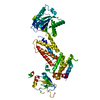
| ||||||||
|---|---|---|---|---|---|---|---|---|---|
| 1 | 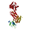
| ||||||||
| Unit cell |
|
-Components
| #1: Protein | Mass: 66528.430 Da / Num. of mol.: 1 Source method: isolated from a genetically manipulated source Source: (gene. exp.) Marinitoga sp. 1155 (bacteria) / Gene: X274_06335 / Production host: |
|---|---|
| Has protein modification | Y |
-Experimental details
-Experiment
| Experiment | Method: X-RAY DIFFRACTION / Number of used crystals: 1 |
|---|
- Sample preparation
| Crystal | Density Matthews: 3.41 Å3/Da / Density % sol: 63.95 % |
|---|---|
| Crystal grow | Temperature: 298 K / Method: vapor diffusion, hanging drop Details: 0.1 M Sodium acetate trihydrate pH 4.6, 8% w/v Polyethylene glycol 4000 |
-Data collection
| Diffraction | Mean temperature: 80 K / Serial crystal experiment: N |
|---|---|
| Diffraction source | Source: SYNCHROTRON / Site: SSRF / Beamline: BL02U1 / Wavelength: 0.979 Å |
| Detector | Type: DECTRIS PILATUS 6M / Detector: PIXEL / Date: Oct 14, 2023 |
| Radiation | Protocol: SINGLE WAVELENGTH / Monochromatic (M) / Laue (L): M / Scattering type: x-ray |
| Radiation wavelength | Wavelength: 0.979 Å / Relative weight: 1 |
| Reflection | Resolution: 3.65→38.39 Å / Num. obs: 10761 / % possible obs: 99.8 % / Redundancy: 34.1 % / Rmerge(I) obs: 0.153 / Net I/σ(I): 22.3 |
| Reflection shell | Resolution: 3.65→3.78 Å / Num. unique obs: 1046 / CC1/2: 0.955 |
- Processing
| Software |
| |||||||||||||||||||||||||||||||||||||||||||||||||||||||||||||||||||||||||||
|---|---|---|---|---|---|---|---|---|---|---|---|---|---|---|---|---|---|---|---|---|---|---|---|---|---|---|---|---|---|---|---|---|---|---|---|---|---|---|---|---|---|---|---|---|---|---|---|---|---|---|---|---|---|---|---|---|---|---|---|---|---|---|---|---|---|---|---|---|---|---|---|---|---|---|---|---|
| Refinement | Method to determine structure: MOLECULAR REPLACEMENT / Resolution: 3.65→38.39 Å / SU ML: 0.34 / Cross valid method: THROUGHOUT / σ(F): 1.35 / Phase error: 25.29 / Stereochemistry target values: ML
| |||||||||||||||||||||||||||||||||||||||||||||||||||||||||||||||||||||||||||
| Solvent computation | Shrinkage radii: 0.9 Å / VDW probe radii: 1.1 Å / Solvent model: FLAT BULK SOLVENT MODEL | |||||||||||||||||||||||||||||||||||||||||||||||||||||||||||||||||||||||||||
| Refinement step | Cycle: LAST / Resolution: 3.65→38.39 Å
| |||||||||||||||||||||||||||||||||||||||||||||||||||||||||||||||||||||||||||
| Refine LS restraints |
| |||||||||||||||||||||||||||||||||||||||||||||||||||||||||||||||||||||||||||
| LS refinement shell |
| |||||||||||||||||||||||||||||||||||||||||||||||||||||||||||||||||||||||||||
| Refinement TLS params. | Method: refined / Refine-ID: X-RAY DIFFRACTION
| |||||||||||||||||||||||||||||||||||||||||||||||||||||||||||||||||||||||||||
| Refinement TLS group |
|
