 Movie
Movie Controller

[English] 日本語
 Yorodumi
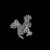
- PDB-8xxu: Cryo-EM Structure of the Prostaglandin D2 Receptor 2 Coupled to G... -
+ Open data

- Basic information
| Entry | Database: PDB / ID: 8xxu | ||||||||||||||||||||||||
|---|---|---|---|---|---|---|---|---|---|---|---|---|---|---|---|---|---|---|---|---|---|---|---|---|---|
| Title | Cryo-EM Structure of the Prostaglandin D2 Receptor 2 Coupled to G Protein | ||||||||||||||||||||||||
 Components Components |
| ||||||||||||||||||||||||
 Keywords Keywords | MEMBRANE PROTEIN / GPCR / Prostaglandin / PTGDR2 | ||||||||||||||||||||||||
| Function / homology |  Function and homology information Function and homology informationprostaglandin J receptor activity / prostaglandin D receptor activity / prostaglandin F receptor activity / negative regulation of male germ cell proliferation / Prostanoid ligand receptors / cellular response to prostaglandin D stimulus / positive regulation of G protein-coupled receptor signaling pathway / neuropeptide binding / adenylate cyclase inhibitor activity / positive regulation of protein localization to cell cortex ...prostaglandin J receptor activity / prostaglandin D receptor activity / prostaglandin F receptor activity / negative regulation of male germ cell proliferation / Prostanoid ligand receptors / cellular response to prostaglandin D stimulus / positive regulation of G protein-coupled receptor signaling pathway / neuropeptide binding / adenylate cyclase inhibitor activity / positive regulation of protein localization to cell cortex / T cell migration / positive regulation of relaxation of smooth muscle / Adenylate cyclase inhibitory pathway / D2 dopamine receptor binding / adenylate cyclase-inhibiting serotonin receptor signaling pathway / G protein-coupled serotonin receptor binding / cellular response to forskolin / regulation of mitotic spindle organization / chemokine-mediated signaling pathway / Regulation of insulin secretion / neuropeptide signaling pathway / calcium-mediated signaling / response to prostaglandin E / positive regulation of cholesterol biosynthetic process / negative regulation of insulin secretion / G protein-coupled receptor binding / response to peptide hormone / G protein-coupled receptor activity / chemotaxis / centriolar satellite / G-protein beta/gamma-subunit complex binding / adenylate cyclase-modulating G protein-coupled receptor signaling pathway / adenylate cyclase-inhibiting G protein-coupled receptor signaling pathway / Olfactory Signaling Pathway / Activation of the phototransduction cascade / G protein-coupled acetylcholine receptor signaling pathway / G beta:gamma signalling through PLC beta / Presynaptic function of Kainate receptors / Thromboxane signalling through TP receptor / Activation of G protein gated Potassium channels / Inhibition of voltage gated Ca2+ channels via Gbeta/gamma subunits / G-protein activation / Glucagon signaling in metabolic regulation / Prostacyclin signalling through prostacyclin receptor / G beta:gamma signalling through CDC42 / Synthesis, secretion, and inactivation of Glucagon-like Peptide-1 (GLP-1) / G beta:gamma signalling through BTK / photoreceptor disc membrane / ADP signalling through P2Y purinoceptor 12 / Glucagon-type ligand receptors / GDP binding / Sensory perception of sweet, bitter, and umami (glutamate) taste / Adrenaline,noradrenaline inhibits insulin secretion / Vasopressin regulates renal water homeostasis via Aquaporins / Glucagon-like Peptide-1 (GLP1) regulates insulin secretion / G alpha (z) signalling events / cellular response to catecholamine stimulus / ADP signalling through P2Y purinoceptor 1 / ADORA2B mediated anti-inflammatory cytokines production / G beta:gamma signalling through PI3Kgamma / adenylate cyclase-activating dopamine receptor signaling pathway / Cooperation of PDCL (PhLP1) and TRiC/CCT in G-protein beta folding / GPER1 signaling / cellular response to prostaglandin E stimulus / heterotrimeric G-protein complex / G alpha (12/13) signalling events / G-protein beta-subunit binding / Inactivation, recovery and regulation of the phototransduction cascade / extracellular vesicle / sensory perception of taste / sperm principal piece / Thrombin signalling through proteinase activated receptors (PARs) / adenylate cyclase-activating G protein-coupled receptor signaling pathway / signaling receptor complex adaptor activity / retina development in camera-type eye / GTPase binding / fibroblast proliferation / G protein activity / midbody / Ca2+ pathway / cell cortex / High laminar flow shear stress activates signaling by PIEZO1 and PECAM1:CDH5:KDR in endothelial cells / G alpha (i) signalling events / G alpha (s) signalling events / phospholipase C-activating G protein-coupled receptor signaling pathway / G alpha (q) signalling events / Hydrolases; Acting on acid anhydrides; Acting on GTP to facilitate cellular and subcellular movement / Ras protein signal transduction / Extra-nuclear estrogen signaling / cell population proliferation / neuron projection / immune response / ciliary basal body / G protein-coupled receptor signaling pathway / cell division / lysosomal membrane / GTPase activity / centrosome / synapse / GTP binding Similarity search - Function | ||||||||||||||||||||||||
| Biological species |  Homo sapiens (human) Homo sapiens (human) | ||||||||||||||||||||||||
| Method | ELECTRON MICROSCOPY / single particle reconstruction / cryo EM / Resolution: 2.54 Å | ||||||||||||||||||||||||
 Authors Authors | Xu, J. / Xu, Y. / Wu, C. / Xu, H.E. | ||||||||||||||||||||||||
| Funding support |  China, 1items China, 1items
| ||||||||||||||||||||||||
 Citation Citation | Journal: Proc Natl Acad Sci U S A / Year: 2024 Title: Molecular basis of lipid and ligand regulation of prostaglandin receptor DP2. Authors: Jiuyin Xu / Youwei Xu / Li Hou / Xinheng He / Yang Li / Jing Zhao / Xue Meng / James Jiqi Wang / Yanli Wu / Heng Zhang / Yunhai Li / Wen Hu / Qingning Yuan / Kai Wu / Xi Cheng / Yi Jiang / ...Authors: Jiuyin Xu / Youwei Xu / Li Hou / Xinheng He / Yang Li / Jing Zhao / Xue Meng / James Jiqi Wang / Yanli Wu / Heng Zhang / Yunhai Li / Wen Hu / Qingning Yuan / Kai Wu / Xi Cheng / Yi Jiang / Yu Xia / H Eric Xu / Canrong Wu / Abstract: Prostaglandin D2 receptor 2 (DP2) is an important anti-inflammatory and antiallergic drug target. While inactive DP2 structures are known, its activation mechanisms and biased signaling remain ...Prostaglandin D2 receptor 2 (DP2) is an important anti-inflammatory and antiallergic drug target. While inactive DP2 structures are known, its activation mechanisms and biased signaling remain unclear. Here, we report cryo-EM structures of an apo DP2-Gi complex, a DP2-Gi complex bound to the endogenous ligand Prostaglandin D (PGD), and a DP2-Gi complex bound to indomethacin, an arrestin-biased ligand, at resolutions of 2.5 Å, 2.8Å, and 2.3 Å, respectively. These structures reveal a distinct binding pose of PGD and indomethacin and provide key insights into receptor activation and transducer coupling. Combining the structural data with functional studies, we uncover the molecular basis for biased signaling of indomethacin toward β-arrestin over G proteins. Notably, a phospholipid binding site was identified at the DP2-G protein interface that modulates DP2-G protein interactions. Together, our functional and structural findings provide insights into DP2 activation, biased signaling, drug interactions, and lipid regulation, enabling rational design of safer antiallergy therapeutics targeting this key immune receptor. | ||||||||||||||||||||||||
| History |
|
- Structure visualization
| Structure viewer | Molecule: MolmilJmol/JSmol |
|---|
- Downloads & links
-Download
| PDBx/mmCIF format | 8xxu.cif.gz | 219.9 KB | Display | PDBx/mmCIF format |
|---|---|---|---|---|
| PDB format | pdb8xxu.ent.gz | Display | PDB format | |
| PDBx/mmJSON format | 8xxu.json.gz | Tree view | PDBx/mmJSON format | |
| Others |  Other downloads Other downloads |
-Validation report
| Arichive directory | https://data.pdbj.org/pub/pdb/validation_reports/xx/8xxuftp://data.pdbj.org/pub/pdb/validation_reports/xx/8xxu | HTTPS FTP |
|---|
-Related structure data
| Related structure data |  38761MC  8xxvC  9iybC M: map data used to model this data C: citing same article ( |
|---|---|
| Similar structure data |

-Links
 PDBj
PDBj


- Assembly
| Deposited unit | 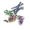
|
|---|---|
| 1 |
|
-Components
-Guanine nucleotide-binding protein ... , 3 types, 3 molecules BCD
| #2: Protein | Mass: 40414.047 Da / Num. of mol.: 1 / Mutation: S47N, G203A, E245A, A326S Source method: isolated from a genetically manipulated source Source: (gene. exp.) Homo sapiens (human) / Gene: GNAI1 / Production host:  Trichoplusia ni (cabbage looper) / References: UniProt: P63096 Trichoplusia ni (cabbage looper) / References: UniProt: P63096 |
|---|---|
| #3: Protein | Mass: 37915.496 Da / Num. of mol.: 1 Source method: isolated from a genetically manipulated source Source: (gene. exp.) Homo sapiens (human) / Gene: GNB1 / Production host: Trichoplusia ni (cabbage looper) / References: UniProt: P62873 |
| #4: Protein | Mass: 7861.143 Da / Num. of mol.: 1 Source method: isolated from a genetically manipulated source Source: (gene. exp.) Homo sapiens (human) / Gene: GNG2 / Production host: Trichoplusia ni (cabbage looper) / References: UniProt: P59768 |
-Protein / Antibody / Non-polymers , 3 types, 3 molecules AE
| #1: Protein | Mass: 38340.043 Da / Num. of mol.: 1 Source method: isolated from a genetically manipulated source Source: (gene. exp.) Homo sapiens (human) / Gene: PTGDR2, CRTH2, DL1R, GPR44 / Production host: Trichoplusia ni (cabbage looper) / References: UniProt: Q9Y5Y4 |
|---|---|
| #5: Antibody | Mass: 26408.492 Da / Num. of mol.: 1 Source method: isolated from a genetically manipulated source Source: (gene. exp.) Trichoplusia ni (cabbage looper) |
| #6: Chemical | ChemComp-A1D5Q / [( Mass: 837.069 Da / Num. of mol.: 1 / Source method: obtained synthetically / Formula: C43H81O13P |
-Details
| Has ligand of interest | N |
|---|---|
| Has protein modification | Y |
-Experimental details
-Experiment
| Experiment | Method: ELECTRON MICROSCOPY |
|---|---|
| EM experiment | Aggregation state: PARTICLE / 3D reconstruction method: single particle reconstruction |
- Sample preparation
| Component | Name: PD2R2-G Protein / Type: COMPLEX / Entity ID: #1-#5 / Source: RECOMBINANT |
|---|---|
| Source (natural) | Organism: Homo sapiens (human) |
| Source (recombinant) | Organism: Trichoplusia ni (cabbage looper) |
| Buffer solution | pH: 7.5 |
| Specimen | Embedding applied: NO / Shadowing applied: NO / Staining applied: NO / Vitrification applied: YES |
| Vitrification | Instrument: FEI VITROBOT MARK IV / Cryogen name: ETHANE / Humidity: 100 % |
- Electron microscopy imaging
| Experimental equipment |  Model: Titan Krios / Image courtesy: FEI Company |
|---|---|
| Microscopy | Model: TFS KRIOS |
| Electron gun | Electron source:  FIELD EMISSION GUN / Accelerating voltage: 300 kV / Illumination mode: FLOOD BEAM FIELD EMISSION GUN / Accelerating voltage: 300 kV / Illumination mode: FLOOD BEAM |
| Electron lens | Mode: BRIGHT FIELD / Nominal defocus max: 5000 nm / Nominal defocus min: 1200 nm |
| Image recording | Electron dose: 50 e/Å2 / Film or detector model: FEI FALCON IV (4k x 4k) |
- Processing
| CTF correction | Type: NONE |
|---|---|
| 3D reconstruction | Resolution: 2.54 Å / Resolution method: FSC 0.143 CUT-OFF / Num. of particles: 257025 / Symmetry type: POINT |
