 Movie
Movie Controller

[English] 日本語
 Yorodumi
- PDB-8xm5: Cryo-EM structure of SARS-CoV-2 Omicron EG.5 spike protein(6P), R... -
+ Open data

- Basic information
| Entry | Database: PDB / ID: 8xm5 | ||||||
|---|---|---|---|---|---|---|---|
| Title | Cryo-EM structure of SARS-CoV-2 Omicron EG.5 spike protein(6P), RBD-closed state | ||||||
 Components Components | Spike glycoprotein | ||||||
 Keywords Keywords | VIRAL PROTEIN / SARS-CoV-2 / Omicron / EG.5 / spike protein | ||||||
| Function / homology |  Function and homology information Function and homology informationsymbiont-mediated disruption of host tissue / Maturation of spike protein / Translation of Structural Proteins / Virion Assembly and Release / host cell surface / host extracellular region / symbiont-mediated-mediated suppression of host tetherin activity / Induction of Cell-Cell Fusion / structural constituent of virion / positive regulation of viral entry into host cell ...symbiont-mediated disruption of host tissue / Maturation of spike protein / Translation of Structural Proteins / Virion Assembly and Release / host cell surface / host extracellular region / symbiont-mediated-mediated suppression of host tetherin activity / Induction of Cell-Cell Fusion / structural constituent of virion / positive regulation of viral entry into host cell / membrane fusion / host cell endoplasmic reticulum-Golgi intermediate compartment membrane / Attachment and Entry / entry receptor-mediated virion attachment to host cell / receptor-mediated virion attachment to host cell / host cell surface receptor binding / symbiont-mediated suppression of host innate immune response / endocytosis involved in viral entry into host cell / receptor ligand activity / fusion of virus membrane with host plasma membrane / fusion of virus membrane with host endosome membrane / viral envelope / symbiont entry into host cell / virion attachment to host cell / host cell plasma membrane / SARS-CoV-2 activates/modulates innate and adaptive immune responses / virion membrane / membrane / identical protein binding / plasma membrane Similarity search - Function | ||||||
| Biological species |   Severe acute respiratory syndrome coronavirus 2 Severe acute respiratory syndrome coronavirus 2 | ||||||
| Method | ELECTRON MICROSCOPY / single particle reconstruction / cryo EM / Resolution: 2.61 Å | ||||||
 Authors Authors | Li, L.J. / Gu, Y.H. / Shi, K.Y. / Qi, J.X. / Gao, G.F. | ||||||
| Funding support |  China, 1items China, 1items
| ||||||
 Citation Citation | Journal: Structure / Year: 2024 Title: Spike structures, receptor binding, and immune escape of recently circulating SARS-CoV-2 Omicron BA.2.86, JN.1, EG.5, EG.5.1, and HV.1 sub-variants. Authors: Linjie Li / Kaiyuan Shi / Yuhang Gu / Zepeng Xu / Chang Shu / Dedong Li / Junqing Sun / Mengqing Cong / Xiaomei Li / Xin Zhao / Guanghui Yu / Songnian Hu / Hui Tan / Jianxun Qi / Xiaopeng Ma ...Authors: Linjie Li / Kaiyuan Shi / Yuhang Gu / Zepeng Xu / Chang Shu / Dedong Li / Junqing Sun / Mengqing Cong / Xiaomei Li / Xin Zhao / Guanghui Yu / Songnian Hu / Hui Tan / Jianxun Qi / Xiaopeng Ma / Kefang Liu / George F Gao / Abstract: The recently emerged BA.2.86, JN.1, EG.5, EG.5.1, and HV.1 variants have a growth advantage. In this study, we explore the structural bases of receptor binding and immune evasion for the Omicron BA.2. ...The recently emerged BA.2.86, JN.1, EG.5, EG.5.1, and HV.1 variants have a growth advantage. In this study, we explore the structural bases of receptor binding and immune evasion for the Omicron BA.2.86, JN.1, EG.5, EG.5.1, and HV.1 sub-variants. Our findings reveal that BA.2.86 exhibits strong receptor binding, whereas its JN.1 sub-lineage displays a decreased binding affinity to human ACE2 (hACE2). Through complex structure analyses, we observed that the reversion of R493Q in BA.2.86 receptor binding domain (RBD) plays a facilitating role in receptor binding, while the L455S substitution in JN.1 RBD restores optimal affinity. Furthermore, the structure of monoclonal antibody (mAb) S309 complexed with BA.2.86 RBD highlights the importance of the K356T mutation, which brings a new N-glycosylation motif, altering the binding pattern of mAbs belonging to RBD-5 represented by S309. These findings emphasize the importance of closely monitoring BA.2.86 and its sub-lineages to prevent another wave of SARS-CoV-2 infections. | ||||||
| History |
|
- Structure visualization
| Structure viewer | Molecule: MolmilJmol/JSmol |
|---|
- Downloads & links
-Download
| PDBx/mmCIF format | 8xm5.cif.gz | 620.1 KB | Display | PDBx/mmCIF format |
|---|---|---|---|---|
| PDB format | pdb8xm5.ent.gz | 492.4 KB | Display | PDB format |
| PDBx/mmJSON format | 8xm5.json.gz | Tree view | PDBx/mmJSON format | |
| Others |  Other downloads Other downloads |
-Validation report
| Arichive directory | https://data.pdbj.org/pub/pdb/validation_reports/xm/8xm5ftp://data.pdbj.org/pub/pdb/validation_reports/xm/8xm5 | HTTPS FTP |
|---|
-Related structure data
| Related structure data |  38463MC 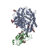 8wp8C  8xlvC 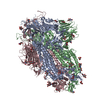 8xmgC 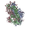 8xmtC 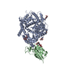 8xn2C 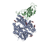 8xn3C 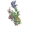 8xn5C 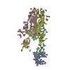 8xnfC 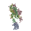 8xnkC 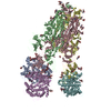 8y16C  8y18C 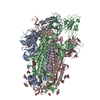 8y5jC 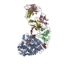 8y6aC M: map data used to model this data C: citing same article ( |
|---|---|
| Similar structure data |


-Links
 PDBj
PDBj


- Assembly
| Deposited unit | 
|
|---|---|
| 1 |
|
-Components
| #1: Protein | Mass: 141066.750 Da / Num. of mol.: 3 / Mutation: F817P, A892P, A899P, A942P, K986P, V987P Source method: isolated from a genetically manipulated source Details: Omicron EG.5 Source: (gene. exp.) Severe acute respiratory syndrome coronavirus 2Gene: S, 2 / Production host:  Homo sapiens (human) / References: UniProt: P0DTC2 Homo sapiens (human) / References: UniProt: P0DTC2#2: Sugar | ChemComp-NAG /   Type: D-saccharide, beta linking / Mass: 221.208 Da / Num. of mol.: 27 / Source method: obtained synthetically / Formula: C8H15NO6 / Feature type: SUBJECT OF INVESTIGATION Type: D-saccharide, beta linking / Mass: 221.208 Da / Num. of mol.: 27 / Source method: obtained synthetically / Formula: C8H15NO6 / Feature type: SUBJECT OF INVESTIGATIONHas ligand of interest | Y | Has protein modification | Y | |
|---|
-Experimental details
-Experiment
| Experiment | Method: ELECTRON MICROSCOPY |
|---|---|
| EM experiment | Aggregation state: PARTICLE / 3D reconstruction method: single particle reconstruction |
- Sample preparation
| Component | Name: SARS-CoV-2 Omicron EG.5 spike protein(6P), RBD-closed state Type: COMPLEX / Entity ID: #1 / Source: RECOMBINANT |
|---|---|
| Source (natural) | Organism: Severe acute respiratory syndrome coronavirus 2 |
| Source (recombinant) | Organism: Homo sapiens (human) |
| Buffer solution | pH: 8 |
| Specimen | Embedding applied: NO / Shadowing applied: NO / Staining applied: NO / Vitrification applied: YES |
| Vitrification | Cryogen name: ETHANE |
- Electron microscopy imaging
| Experimental equipment |  Model: Titan Krios / Image courtesy: FEI Company |
|---|---|
| Microscopy | Model: FEI TITAN KRIOS |
| Electron gun | Electron source:  FIELD EMISSION GUN / Accelerating voltage: 300 kV / Illumination mode: FLOOD BEAM FIELD EMISSION GUN / Accelerating voltage: 300 kV / Illumination mode: FLOOD BEAM |
| Electron lens | Mode: BRIGHT FIELD / Nominal defocus max: 2000 nm / Nominal defocus min: 1000 nm |
| Image recording | Electron dose: 50 e/Å2 / Film or detector model: FEI FALCON IV (4k x 4k) |
- Processing
| EM software | Name: PHENIX / Version: 1.20.1_4487: / Category: model refinement | ||||||||||||||||||||||||
|---|---|---|---|---|---|---|---|---|---|---|---|---|---|---|---|---|---|---|---|---|---|---|---|---|---|
| CTF correction | Type: PHASE FLIPPING AND AMPLITUDE CORRECTION | ||||||||||||||||||||||||
| 3D reconstruction | Resolution: 2.61 Å / Resolution method: FSC 0.143 CUT-OFF / Num. of particles: 473611 / Symmetry type: POINT | ||||||||||||||||||||||||
| Refine LS restraints |
|
