 Movie
Movie Controller

[English] 日本語
 Yorodumi
- EMDB-38460: Cryo-EM structure of SARS-CoV-2 Omicron BA.2.86 spike protein(6P)... -
+ Open data

- Basic information
| Entry |  | |||||||||
|---|---|---|---|---|---|---|---|---|---|---|
| Title | Cryo-EM structure of SARS-CoV-2 Omicron BA.2.86 spike protein(6P), 1-RBD-up state | |||||||||
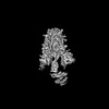
 Map data Map data | Cryo-EM map(sharp) of SARS-CoV-2 Omicron BA.2.86 spike protein | |||||||||
 Sample Sample |
| |||||||||
 Keywords Keywords | SARS-CoV-2 / Omicron / BA.2.86 / spike protein / VIRAL PROTEIN | |||||||||
| Function / homology |  Function and homology information Function and homology informationsymbiont-mediated disruption of host tissue / Maturation of spike protein / Translation of Structural Proteins / Virion Assembly and Release / host cell surface / host extracellular region / symbiont-mediated-mediated suppression of host tetherin activity / Induction of Cell-Cell Fusion / structural constituent of virion / positive regulation of viral entry into host cell ...symbiont-mediated disruption of host tissue / Maturation of spike protein / Translation of Structural Proteins / Virion Assembly and Release / host cell surface / host extracellular region / symbiont-mediated-mediated suppression of host tetherin activity / Induction of Cell-Cell Fusion / structural constituent of virion / positive regulation of viral entry into host cell / membrane fusion / host cell endoplasmic reticulum-Golgi intermediate compartment membrane / Attachment and Entry / entry receptor-mediated virion attachment to host cell / receptor-mediated virion attachment to host cell / host cell surface receptor binding / symbiont-mediated suppression of host innate immune response / endocytosis involved in viral entry into host cell / receptor ligand activity / fusion of virus membrane with host plasma membrane / fusion of virus membrane with host endosome membrane / viral envelope / symbiont entry into host cell / virion attachment to host cell / host cell plasma membrane / SARS-CoV-2 activates/modulates innate and adaptive immune responses / virion membrane / membrane / identical protein binding / plasma membrane Similarity search - Function | |||||||||
| Biological species |   Severe acute respiratory syndrome coronavirus 2 Severe acute respiratory syndrome coronavirus 2 | |||||||||
| Method | single particle reconstruction / cryo EM / Resolution: 3.07 Å | |||||||||
 Authors Authors | Li LJ / Gu YH / Shi KY / Qi JX / Gao GF | |||||||||
| Funding support |  China, 1 items China, 1 items
| |||||||||
 Citation Citation | Journal: Structure / Year: 2024 Title: Spike structures, receptor binding, and immune escape of recently circulating SARS-CoV-2 Omicron BA.2.86, JN.1, EG.5, EG.5.1, and HV.1 sub-variants. Authors: Linjie Li / Kaiyuan Shi / Yuhang Gu / Zepeng Xu / Chang Shu / Dedong Li / Junqing Sun / Mengqing Cong / Xiaomei Li / Xin Zhao / Guanghui Yu / Songnian Hu / Hui Tan / Jianxun Qi / Xiaopeng Ma ...Authors: Linjie Li / Kaiyuan Shi / Yuhang Gu / Zepeng Xu / Chang Shu / Dedong Li / Junqing Sun / Mengqing Cong / Xiaomei Li / Xin Zhao / Guanghui Yu / Songnian Hu / Hui Tan / Jianxun Qi / Xiaopeng Ma / Kefang Liu / George F Gao / Abstract: The recently emerged BA.2.86, JN.1, EG.5, EG.5.1, and HV.1 variants have a growth advantage. In this study, we explore the structural bases of receptor binding and immune evasion for the Omicron BA.2. ...The recently emerged BA.2.86, JN.1, EG.5, EG.5.1, and HV.1 variants have a growth advantage. In this study, we explore the structural bases of receptor binding and immune evasion for the Omicron BA.2.86, JN.1, EG.5, EG.5.1, and HV.1 sub-variants. Our findings reveal that BA.2.86 exhibits strong receptor binding, whereas its JN.1 sub-lineage displays a decreased binding affinity to human ACE2 (hACE2). Through complex structure analyses, we observed that the reversion of R493Q in BA.2.86 receptor binding domain (RBD) plays a facilitating role in receptor binding, while the L455S substitution in JN.1 RBD restores optimal affinity. Furthermore, the structure of monoclonal antibody (mAb) S309 complexed with BA.2.86 RBD highlights the importance of the K356T mutation, which brings a new N-glycosylation motif, altering the binding pattern of mAbs belonging to RBD-5 represented by S309. These findings emphasize the importance of closely monitoring BA.2.86 and its sub-lineages to prevent another wave of SARS-CoV-2 infections. | |||||||||
| History |
|
- Structure visualization
| Supplemental images |
|---|

- Downloads & links
-EMDB archive
| Map data | emd_38460.map.gz | 421.4 MB | EMDB map data format | |
|---|---|---|---|---|
| Header (meta data) | emd-38460-v30.xmlemd-38460.xml | 15.6 KB 15.6 KB | Display Display | EMDB header |
| Images |  emd_38460.png emd_38460.png | 100.3 KB | ||
| Masks | emd_38460_msk_1.map | 476.8 MB | Mask map | |
| Filedesc metadata | emd-38460.cif.gz | 6.3 KB | ||
| Others | emd_38460_half_map_1.map.gzemd_38460_half_map_2.map.gz | 442.8 MB 442.8 MB | ||
| Archive directory |  http://ftp.pdbj.org/pub/emdb/structures/EMD-38460ftp://ftp.pdbj.org/pub/emdb/structures/EMD-38460 http://ftp.pdbj.org/pub/emdb/structures/EMD-38460ftp://ftp.pdbj.org/pub/emdb/structures/EMD-38460 | HTTPS FTP |
-Related structure data
| Related structure data |  8xlvMC 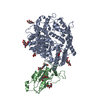 8wp8C  8xm5C 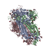 8xmgC 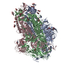 8xmtC 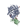 8xn2C 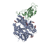 8xn3C 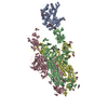 8xn5C 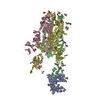 8xnfC 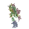 8xnkC 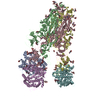 8y16C  8y18C 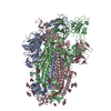 8y5jC 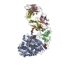 8y6aC M: atomic model generated by this map C: citing same article ( |
|---|---|
| Similar structure data |


-Links
| EMDB pages | EMDB (EBI/PDBe) / EMDataResource |
|---|---|
| Related items in Molecule of the Month |


-Map
| File | Download / File: emd_38460.map.gz / Format: CCP4 / Size: 476.8 MB / Type: IMAGE STORED AS FLOATING POINT NUMBER (4 BYTES) | ||||||||||||||||||||||||||||||||||||
|---|---|---|---|---|---|---|---|---|---|---|---|---|---|---|---|---|---|---|---|---|---|---|---|---|---|---|---|---|---|---|---|---|---|---|---|---|---|
| Annotation | Cryo-EM map(sharp) of SARS-CoV-2 Omicron BA.2.86 spike protein | ||||||||||||||||||||||||||||||||||||
| Projections & slices | Image control
Images are generated by Spider. | ||||||||||||||||||||||||||||||||||||
| Voxel size | X=Y=Z: 0.669 Å | ||||||||||||||||||||||||||||||||||||
| Density |
| ||||||||||||||||||||||||||||||||||||
| Symmetry | Space group: 1 | ||||||||||||||||||||||||||||||||||||
| Details | EMDB XML:
|
 Z (Sec.)
 Y (Row.)
 X (Col.)


-Supplemental data
-Mask #1
| File | emd_38460_msk_1.map | ||||||||||||
|---|---|---|---|---|---|---|---|---|---|---|---|---|---|
| Projections & Slices |
| ||||||||||||
| Density Histograms |


-Half map: Half-B map of SARS-CoV-2 Omicron BA.2.86 spike protein
| File | emd_38460_half_map_1.map | ||||||||||||
|---|---|---|---|---|---|---|---|---|---|---|---|---|---|
| Annotation | Half-B map of SARS-CoV-2 Omicron BA.2.86 spike protein | ||||||||||||
| Projections & Slices |
| ||||||||||||
| Density Histograms |


-Half map: Half-A map of SARS-CoV-2 Omicron BA.2.86 spike protein
| File | emd_38460_half_map_2.map | ||||||||||||
|---|---|---|---|---|---|---|---|---|---|---|---|---|---|
| Annotation | Half-A map of SARS-CoV-2 Omicron BA.2.86 spike protein | ||||||||||||
| Projections & Slices |
| ||||||||||||
| Density Histograms |


- Sample components
-Entire : SARS-CoV-2 Omicron BA.2.86 spike protein
| Entire | Name: SARS-CoV-2 Omicron BA.2.86 spike protein |
|---|---|
| Components |
|
-Supramolecule #1: SARS-CoV-2 Omicron BA.2.86 spike protein
| Supramolecule | Name: SARS-CoV-2 Omicron BA.2.86 spike protein / type: complex / ID: 1 / Parent: 0 / Macromolecule list: #1 |
|---|---|
| Source (natural) | Organism: Severe acute respiratory syndrome coronavirus 2 |
-Macromolecule #1: Spike glycoprotein
| Macromolecule | Name: Spike glycoprotein / type: protein_or_peptide / ID: 1 / Details: Omicron BA.2.86 / Number of copies: 3 / Enantiomer: LEVO |
|---|---|
| Source (natural) | Organism: Severe acute respiratory syndrome coronavirus 2 |
| Molecular weight | Theoretical: 131.772406 KDa |
| Recombinant expression | Organism:  Homo sapiens (human) Homo sapiens (human) |
| Sequence | String: MFVFLVLLPL VSSQCVMPLF NLITTTQSYT NSFTRGVYYP DKVFRSSVLH LTQDLFLPFF SNVTWFHAIS GTNGTKRFDN PVLPFNDGV YFASTEKSNI IRGWIFGTTL DSKTQSLLIV NNATNVFIKV CEFQFCNDPF LDVYHKNNKS WMESESGVYS S ANNCTFEY ...String: MFVFLVLLPL VSSQCVMPLF NLITTTQSYT NSFTRGVYYP DKVFRSSVLH LTQDLFLPFF SNVTWFHAIS GTNGTKRFDN PVLPFNDGV YFASTEKSNI IRGWIFGTTL DSKTQSLLIV NNATNVFIKV CEFQFCNDPF LDVYHKNNKS WMESESGVYS S ANNCTFEY VSQPFLMDLE GKQGNFKNLR EFVFKNIDGY FKIYSKHTPI IGRDFPQGFS ALEPLVDLPI GINITRFQTL LA LNRSYLT PGDSSSGWTA GAADYYVGYL QPRTFLLKYN ENGTITDAVD CALDPLSETK CTLKSFTVEK GIYQTSNFRV QPT ESIVRF PNVTNLCPFH EVFNATRFAS VYAWNRTRIS NCVADYSVLY NFAPFFAFKC YGVSPTKLND LCFTNVYADS FVIK GNEVS QIAPGQTGNI ADYNYKLPDD FTGCVIAWNS NKLDSKHSGN YDYWYRLFRK SKLKPFERDI STEIYQAGNK PCKGK GPNC YFPLQSYGFR PTYGVGHQPY RVVVLSFELL HAPATVCGPK KSTNLVKNKC VNFNFNGLTG TGVLTKSNKK FLPFQQ FGR DIVDTTDAVR DPQTLEILDI TPCSFGGVSV ITPGTNTSNQ VAVLYQGVNC TEVSVAIHAD QLTPTWRVYS TGSNVFQ TR AGCLIGAEYV NNSYECDIPI GAGVCASYQT QTKSRGSASS VASQSIIAYT MSLGAENSVA YSNNSIAIPT NFTISVTT E ILPVSMTKTS VDCTMYICGD STECSNLLLQ YGSFCTQLKR ALTGIAVEQD KNTQEVFAQV KQIYKTPPIK YFGGFNFSQ ILPDPSKPSK RSPIEDLLFN KVTLADAGFI KQYGDCLGDI AARDLICAQK FNGLTVLPPL LTDEMIAQYT SALLAGTITS GWTFGAGPA LQIPFPMQMA YRFNGIGVTQ NVLYENQKLI ANQFNSAIGK IQDSLFSTPS ALGKLQDVVN HNAQALNTLV K QLSSKFGA ISSVLNDILS RLDPPEAEVQ IDRLITGRLQ SLQTYVTQQL IRAAEIRASA NLAATKMSEC VLGQSKRVDF CG KGYHLMS FPQSAPHGVV FLHVTYVPAQ EKNFTTAPAI CHDGKAHFPR EGVFVSNGTH WFVTQRNFYE PQIITTDNTF VSG NCDVVI GIVNNTVYDP LQLELDSGGG SGGGSGYIPE APRDGQAYVR KDGEWVLLST FLGGGSAWSH PQFEK UniProtKB: Spike glycoprotein |
-Macromolecule #3: 2-acetamido-2-deoxy-beta-D-glucopyranose
| Macromolecule | Name: 2-acetamido-2-deoxy-beta-D-glucopyranose / type: ligand / ID: 3 / Number of copies: 24 / Formula: NAG |
|---|---|
| Molecular weight | Theoretical: 221.208 Da |
| Chemical component information |  ChemComp-NAG: |
-Experimental details
-Structure determination
| Method | cryo EM |
|---|---|
 Processing Processing | single particle reconstruction |
| Aggregation state | particle |
-Sample preparation
| Buffer | pH: 8 |
|---|---|
| Vitrification | Cryogen name: ETHANE |
- Electron microscopy
| Microscope | FEI TITAN KRIOS |
|---|---|
| Image recording | Film or detector model: GATAN K3 (6k x 4k) / Average electron dose: 60.0 e/Å2 |
| Electron beam | Acceleration voltage: 300 kV / Electron source:  FIELD EMISSION GUN FIELD EMISSION GUN |
| Electron optics | Illumination mode: FLOOD BEAM / Imaging mode: BRIGHT FIELD / Nominal defocus max: 2.0 µm / Nominal defocus min: 1.0 µm |
| Experimental equipment |  Model: Titan Krios / Image courtesy: FEI Company |
-Image processing
| Startup model | Type of model: NONE |
|---|---|
| Final reconstruction | Resolution.type: BY AUTHOR / Resolution: 3.07 Å / Resolution method: FSC 0.143 CUT-OFF / Number images used: 242344 |
| Initial angle assignment | Type: MAXIMUM LIKELIHOOD |
| Final angle assignment | Type: MAXIMUM LIKELIHOOD |
