[English] 日本語
 Yorodumi
- PDB-8xk4: Structure of the Argonaute protein from Kurthia massiliensis in c... -
+ Open data
- Basic information
| Entry | Database: PDB / ID: 8xk4 | ||||||
|---|---|---|---|---|---|---|---|
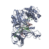
| Title | Structure of the Argonaute protein from Kurthia massiliensis in complex with guide DNA and 19-mer target DNA in target-released state | ||||||
 Components Components |
| ||||||
 Keywords Keywords | DNA BINDING PROTEIN/DNA / Mesophilic prokaryotic Argonaute / DNA BINDING PROTEIN-DNA COMPLEX | ||||||
| Function / homology | 2'-DEOXYCYTIDINE-5'-MONOPHOSPHATE / : / DNA / DNA (> 10) Function and homology information Function and homology information | ||||||
| Biological species |  Kurthia massiliensis (bacteria) Kurthia massiliensis (bacteria) | ||||||
| Method | ELECTRON MICROSCOPY / single particle reconstruction / cryo EM / Resolution: 2.94 Å | ||||||
 Authors Authors | Tao, X. / Ding, H. / Wu, S. | ||||||
| Funding support |  China, 1items China, 1items
| ||||||
 Citation Citation |  Journal: Nucleic Acids Res / Year: 2024 Journal: Nucleic Acids Res / Year: 2024Title: Structural and mechanistic insights into a mesophilic prokaryotic Argonaute. Authors: Xin Tao / Hui Ding / Shaowen Wu / Fei Wang / Hu Xu / Jie Li / Chao Zhai / Shunshun Li / Kai Chen / Shan Wu / Yang Liu / Lixin Ma /  Abstract: Argonaute (Ago) proteins are programmable nucleases found in all domains of life, playing a crucial role in biological processes like DNA/RNA interference and gene regulation. Mesophilic prokaryotic ...Argonaute (Ago) proteins are programmable nucleases found in all domains of life, playing a crucial role in biological processes like DNA/RNA interference and gene regulation. Mesophilic prokaryotic Agos (pAgos) have gained increasing research interest due to their broad range of potential applications, yet their molecular mechanisms remain poorly understood. Here, we present seven cryo-electron microscopy structures of Kurthia massiliensis Ago (KmAgo) in various states. These structures encompass the steps of apo-form, guide binding, target recognition, cleavage, and release, revealing that KmAgo employs a unique DDD catalytic triad, instead of a DEDD tetrad, for DNA target cleavage under 5'P-DNA guide conditions. Notably, the last catalytic residue, D713, is positioned outside the catalytic pocket in the absence of guide. After guide binding, D713 enters the catalytic pocket. In contrast, the corresponding catalytic residue in other Agos has been consistently located in the catalytic pocket. Moreover, we identified several sites exhibiting enhanced catalytic activity through alanine mutagenesis. These sites have the potential to serve as engineering targets for augmenting the catalytic efficiency of KmAgo. This structural analysis of KmAgo advances the understanding of the diversity of molecular mechanisms by Agos, offering insights for developing and optimizing mesophilic pAgos-based programmable DNA and RNA manipulation tools. | ||||||
| History |
|
- Structure visualization
| Structure viewer | Molecule:  Molmil Molmil Jmol/JSmol Jmol/JSmol |
|---|
- Downloads & links
- Download
| PDBx/mmCIF format |  8xk4.cif.gz 8xk4.cif.gz | 197.6 KB | Display |  PDBx/mmCIF format PDBx/mmCIF format |
|---|---|---|---|---|
| PDB format |  pdb8xk4.ent.gz pdb8xk4.ent.gz | 144.1 KB | Display |  PDB format PDB format |
| PDBx/mmJSON format |  8xk4.json.gz 8xk4.json.gz | Tree view |  PDBx/mmJSON format PDBx/mmJSON format | |
| Others |  Other downloads Other downloads |
-Validation report
| Summary document |  8xk4_validation.pdf.gz 8xk4_validation.pdf.gz | 1.2 MB | Display |  wwPDB validaton report wwPDB validaton report |
|---|---|---|---|---|
| Full document |  8xk4_full_validation.pdf.gz 8xk4_full_validation.pdf.gz | 1.2 MB | Display | |
| Data in XML |  8xk4_validation.xml.gz 8xk4_validation.xml.gz | 36.4 KB | Display | |
| Data in CIF |  8xk4_validation.cif.gz 8xk4_validation.cif.gz | 53 KB | Display | |
| Arichive directory |  https://data.pdbj.org/pub/pdb/validation_reports/xk/8xk4 https://data.pdbj.org/pub/pdb/validation_reports/xk/8xk4 ftp://data.pdbj.org/pub/pdb/validation_reports/xk/8xk4 ftp://data.pdbj.org/pub/pdb/validation_reports/xk/8xk4 | HTTPS FTP |
-Related structure data
| Related structure data |  38415MC  8xhsC  8xhvC  8xjwC  8xjxC  8xk0C  8xk3C M: map data used to model this data C: citing same article ( |
|---|---|
| Similar structure data | Similarity search - Function & homology  F&H Search F&H Search |
- Links
Links
- Assembly
| Deposited unit | 
|
|---|---|
| 1 |
|
- Components
-Protein , 1 types, 1 molecules A
| #1: Protein | Mass: 85485.203 Da / Num. of mol.: 1 Source method: isolated from a genetically manipulated source Source: (gene. exp.)  Kurthia massiliensis (bacteria) / Production host: Kurthia massiliensis (bacteria) / Production host:  |
|---|
-DNA chain , 2 types, 2 molecules BC
| #2: DNA chain | Mass: 5641.660 Da / Num. of mol.: 1 / Source method: obtained synthetically / Source: (synth.)  Kurthia massiliensis (bacteria) Kurthia massiliensis (bacteria) |
|---|---|
| #3: DNA chain | Mass: 2963.969 Da / Num. of mol.: 1 / Source method: obtained synthetically / Source: (synth.)  Kurthia massiliensis (bacteria) Kurthia massiliensis (bacteria) |
-Non-polymers , 3 types, 6 molecules 


| #4: Chemical | | #5: Chemical | ChemComp-DC / | #6: Water | ChemComp-HOH / | |
|---|
-Details
| Has ligand of interest | Y |
|---|---|
| Has protein modification | N |
-Experimental details
-Experiment
| Experiment | Method: ELECTRON MICROSCOPY |
|---|---|
| EM experiment | Aggregation state: PARTICLE / 3D reconstruction method: single particle reconstruction |
- Sample preparation
| Component | Name: KmAgo / Type: COMPLEX / Entity ID: #1 / Source: RECOMBINANT |
|---|---|
| Source (natural) | Organism:  Kurthia massiliensis (bacteria) Kurthia massiliensis (bacteria) |
| Source (recombinant) | Organism:  |
| Buffer solution | pH: 7.5 |
| Specimen | Embedding applied: NO / Shadowing applied: NO / Staining applied: NO / Vitrification applied: YES |
| Vitrification | Cryogen name: ETHANE |
- Electron microscopy imaging
| Experimental equipment |  Model: Titan Krios / Image courtesy: FEI Company |
|---|---|
| Microscopy | Model: TFS KRIOS |
| Electron gun | Electron source:  FIELD EMISSION GUN / Accelerating voltage: 300 kV / Illumination mode: FLOOD BEAM FIELD EMISSION GUN / Accelerating voltage: 300 kV / Illumination mode: FLOOD BEAM |
| Electron lens | Mode: BRIGHT FIELD / Nominal defocus max: 1500 nm / Nominal defocus min: 1000 nm |
| Image recording | Electron dose: 54 e/Å2 / Film or detector model: GATAN K3 BIOQUANTUM (6k x 4k) |
- Processing
| CTF correction | Type: PHASE FLIPPING AND AMPLITUDE CORRECTION | ||||||||||||||||||||||||
|---|---|---|---|---|---|---|---|---|---|---|---|---|---|---|---|---|---|---|---|---|---|---|---|---|---|
| 3D reconstruction | Resolution: 2.94 Å / Resolution method: FSC 0.143 CUT-OFF / Num. of particles: 259549 / Symmetry type: POINT | ||||||||||||||||||||||||
| Refinement | Highest resolution: 2.94 Å Stereochemistry target values: REAL-SPACE (WEIGHTED MAP SUM AT ATOM CENTERS) | ||||||||||||||||||||||||
| Refine LS restraints |
|
 Movie
Movie Controller


 PDBj
PDBj


