[English] 日本語
Yorodumi
- PDB-8xi9: Crystal structure of FRB-FKBP fusion protein in complex with rapamycin -

+
Open data


ID or keywords:

Loading...

-
Basic information

Entry
Database: PDB / ID: 8xi9
TitleCrystal structure of FRB-FKBP fusion protein in complex with rapamycin
ComponentsFRB-FKBP fusion protein
KeywordsSIGNALING PROTEIN / Rapamycin
Function / homology
Function and homology information


RNA polymerase III type 2 promoter sequence-specific DNA binding / RNA polymerase III type 1 promoter sequence-specific DNA binding / positive regulation of cytoplasmic translational initiation / T-helper 1 cell lineage commitment / positive regulation of pentose-phosphate shunt / regulation of locomotor rhythm / positive regulation of wound healing, spreading of epidermal cells / cellular response to leucine starvation / TFIIIC-class transcription factor complex binding / TORC2 complex ...RNA polymerase III type 2 promoter sequence-specific DNA binding / RNA polymerase III type 1 promoter sequence-specific DNA binding / positive regulation of cytoplasmic translational initiation / T-helper 1 cell lineage commitment / positive regulation of pentose-phosphate shunt / regulation of locomotor rhythm / positive regulation of wound healing, spreading of epidermal cells / cellular response to leucine starvation / TFIIIC-class transcription factor complex binding / TORC2 complex / regulation of membrane permeability / heart valve morphogenesis / macrolide binding / negative regulation of lysosome organization / activin receptor binding / RNA polymerase III type 3 promoter sequence-specific DNA binding / positive regulation of transcription of nucleolar large rRNA by RNA polymerase I / TORC1 complex / cytoplasmic side of membrane / calcineurin-NFAT signaling cascade / nucleus localization / cellular response to methionine / voluntary musculoskeletal movement / regulation of osteoclast differentiation / regulation of skeletal muscle contraction by regulation of release of sequestered calcium ion / TORC1 signaling / signaling receptor inhibitor activity / positive regulation of keratinocyte migration / transforming growth factor beta receptor binding / TGFBR1 LBD Mutants in Cancer / cellular response to L-leucine / MTOR signalling / Amino acids regulate mTORC1 / type I transforming growth factor beta receptor binding / cellular response to nutrient / regulation of autophagosome assembly / Energy dependent regulation of mTOR by LKB1-AMPK / energy reserve metabolic process / negative regulation of activin receptor signaling pathway / heart trabecula formation / negative regulation of cell size / ruffle organization / terminal cisterna / ryanodine receptor complex / cellular response to osmotic stress / I-SMAD binding / negative regulation of protein localization to nucleus / regulation of amyloid precursor protein catabolic process / anoikis / cardiac muscle cell development / protein maturation by protein folding / positive regulation of transcription by RNA polymerase III / ventricular cardiac muscle tissue morphogenesis / negative regulation of calcineurin-NFAT signaling cascade / 'de novo' protein folding / regulation of myelination / negative regulation of macroautophagy / regulation of cell size / positive regulation of oligodendrocyte differentiation / FK506 binding / Macroautophagy / positive regulation of actin filament polymerization / lysosome organization / positive regulation of myotube differentiation / oligodendrocyte differentiation / behavioral response to pain / channel regulator activity / TGF-beta receptor signaling activates SMADs / Constitutive Signaling by AKT1 E17K in Cancer / germ cell development / mTORC1-mediated signalling / CD28 dependent PI3K/Akt signaling / : / protein peptidyl-prolyl isomerization / Calcineurin activates NFAT / HSF1-dependent transactivation / neuronal action potential / regulation of ryanodine-sensitive calcium-release channel activity / regulation of immune response / TOR signaling / response to amino acid / endomembrane system / cellular response to nutrient levels / positive regulation of translational initiation / regulation of macroautophagy / 'de novo' pyrimidine nucleobase biosynthetic process / positive regulation of lamellipodium assembly / positive regulation of epithelial to mesenchymal transition / positive regulation of lipid biosynthetic process / heart morphogenesis / response to nutrient / cardiac muscle contraction / supramolecular fiber organization / regulation of cellular response to heat / phagocytic vesicle / positive regulation of stress fiber assembly / cytoskeleton organization / Regulation of PTEN gene transcription / sarcoplasmic reticulum membrane / T cell costimulation
Similarity search - Function
Domain of unknown function DUF3385, target of rapamycin protein / Serine/threonine-protein kinase mTOR domain / Domain of unknown function / FKBP12-rapamycin binding domain / Serine/threonine-protein kinase TOR / FKBP12-rapamycin binding domain superfamily / FKBP12-rapamycin binding domain / Rapamycin binding domain / : / FATC domain ...Domain of unknown function DUF3385, target of rapamycin protein / Serine/threonine-protein kinase mTOR domain / Domain of unknown function / FKBP12-rapamycin binding domain / Serine/threonine-protein kinase TOR / FKBP12-rapamycin binding domain superfamily / FKBP12-rapamycin binding domain / Rapamycin binding domain / : / FATC domain / PIK-related kinase, FAT / FAT domain / FATC / FATC domain / PIK-related kinase / FAT domain profile. / FATC domain profile. / : / FKBP-type peptidyl-prolyl cis-trans isomerase domain profile. / FKBP-type peptidyl-prolyl cis-trans isomerase domain / FKBP-type peptidyl-prolyl cis-trans isomerase / Phosphatidylinositol 3- and 4-kinases signature 1. / Phosphatidylinositol 3/4-kinase, conserved site / Phosphatidylinositol 3- and 4-kinases signature 2. / Phosphatidylinositol 3-/4-kinase, catalytic domain superfamily / Phosphoinositide 3-kinase, catalytic domain / Phosphatidylinositol 3- and 4-kinase / Phosphatidylinositol 3- and 4-kinases catalytic domain profile. / Phosphatidylinositol 3-/4-kinase, catalytic domain / Peptidyl-prolyl cis-trans isomerase domain superfamily / Armadillo-like helical / Tetratricopeptide-like helical domain superfamily / Armadillo-type fold / Protein kinase-like domain superfamily
Similarity search - Domain/homology
RAPAMYCIN IMMUNOSUPPRESSANT DRUG / Serine/threonine-protein kinase mTOR / Peptidyl-prolyl cis-trans isomerase FKBP1A
Similarity search - Component
Biological speciesHomo sapiens (human)
MethodX-RAY DIFFRACTION / SYNCHROTRON / MOLECULAR REPLACEMENT / Resolution: 1.85 Å
AuthorsInobe, T. / Sakaguchi, R. / Obita, T. / Mukaiyama, A. / Yokoyama, T. / Mizuguchi, M. / Akiyama, S.
Funding support Japan, 2items
OrganizationGrant numberCountry
Japan Society for the Promotion of Science (JSPS)JP18H04544 Japan
Japan Society for the Promotion of Science (JSPS)JP19K06595 Japan
CitationJournal: Febs Lett. / Year: 2024
Title: Structural insights into rapamycin-induced oligomerization of a FRB-FKBP fusion protein.
Authors: Inobe, T. / Sakaguchi, R. / Obita, T. / Mukaiyama, A. / Koike, S. / Yokoyama, T. / Mizuguchi, M. / Akiyama, S.
History
DepositionDec 19, 2023Deposition site: PDBJ / Processing site: PDBJ
Revision 1.0Aug 7, 2024Provider: repository / Type: Initial release

-
Structure visualization

Structure viewerMolecule:
MolmilJmol/JSmol

Downloads & links

-
Assembly

Deposited unit
A: FRB-FKBP fusion protein
hetero molecules


Theoretical massNumber of molelcules
Total (without water)24,2452
Polymers23,3311
Non-polymers9141
Water2,072115
1


  • Idetical with deposited unit
  • defined by author&software
  • Evidence: SAXS, forms a tetramer or pentamer structure
TypeNameSymmetry operationNumber
identity operation1_555x,y,z1
Unit cell
Length a, b, c (Å)80.517, 80.517, 122.419
Angle α, β, γ (deg.)90.000, 90.000, 120.000
Int Tables number182
Space group name H-MP6322
Space group name HallP6c2c
Symmetry operation#1: x,y,z
#2: x-y,x,z+1/2
#3: y,-x+y,z+1/2
#4: -y,x-y,z
#5: -x+y,-x,z
#6: x-y,-y,-z
#7: -x,-x+y,-z
#8: -x,-y,z+1/2
#9: y,x,-z
#10: -y,-x,-z+1/2
#11: -x+y,y,-z+1/2
#12: x,x-y,-z+1/2
Components on special symmetry positions
IDModelComponents
11A-508-

HOH

-
Components

#1: Protein FRB-FKBP fusion protein


Mass: 23330.688 Da / Num. of mol.: 1
Source method: isolated from a genetically manipulated source
Source: (gene. exp.) Homo sapiens (human)
Gene: MTOR, FRAP, FRAP1, FRAP2, RAFT1, RAPT1, FKBP1A, FKBP1, FKBP12
Production host: Escherichia coli (E. coli)
References: UniProt: P42345, UniProt: P62942, non-specific serine/threonine protein kinase, peptidylprolyl isomerase
#2: Chemical ChemComp-RAP / RAPAMYCIN IMMUNOSUPPRESSANT DRUG


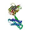
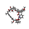
Mass: 914.172 Da / Num. of mol.: 1 / Source method: obtained synthetically / Formula: C51H79NO13 / Feature type: SUBJECT OF INVESTIGATION / Comment: immunosuppressant, antibiotic*YM
#3: Water ChemComp-HOH / water


Mass: 18.015 Da / Num. of mol.: 115 / Source method: isolated from a natural source / Formula: H2O
Has ligand of interestY

-
Experimental details

-
Experiment

ExperimentMethod: X-RAY DIFFRACTION / Number of used crystals: 1

-
Sample preparation

CrystalDensity Matthews: 2.45 Å3/Da / Density % sol: 49.9 %
Crystal growTemperature: 293 K / Method: vapor diffusion, sitting drop / pH: 7.4 / Details: 4.4M Sodium Acetate

-
Data collection

DiffractionMean temperature: 100 K / Serial crystal experiment: N
Diffraction sourceSource: SYNCHROTRON / Site: SLS / Beamline: X06SA / Wavelength: 1 Å
DetectorType: DECTRIS EIGER X 16M / Detector: PIXEL / Date: Aug 27, 2021
RadiationProtocol: SINGLE WAVELENGTH / Monochromatic (M) / Laue (L): M / Scattering type: x-ray
Radiation wavelengthWavelength: 1 Å / Relative weight: 1
ReflectionResolution: 1.85→69.73 Å / Num. obs: 20689 / % possible obs: 99.9 % / Redundancy: 37.3 % / Biso Wilson estimate: 25.31 Å2 / CC1/2: 1 / Rmerge(I) obs: 0.059 / Rpim(I) all: 0.013 / Rrim(I) all: 0.06 / Net I/σ(I): 44.9
Reflection shellResolution: 1.85→1.89 Å / Rmerge(I) obs: 0.335 / Mean I/σ(I) obs: 13.2 / Num. unique obs: 1239 / CC1/2: 0.992 / Rpim(I) all: 0.075 / Rrim(I) all: 0.343

-
Processing

Software
NameVersionClassification
PHENIX1.20.1_4487refinement
Cootmodel building
MOSFLMdata reduction
Aimlessdata scaling
PHASERphasing
RefinementMethod to determine structure: MOLECULAR REPLACEMENT / Resolution: 1.85→61.21 Å / SU ML: 0.1519 / Cross valid method: FREE R-VALUE / σ(F): 1.34 / Phase error: 21.2749
Stereochemistry target values: GeoStd + Monomer Library + CDL v1.2
RfactorNum. reflection% reflection
Rfree0.2184 1053 5.1 %
Rwork0.1872 19596 -
obs0.1887 20649 99.84 %
Solvent computationShrinkage radii: 0.9 Å / VDW probe radii: 1.1 Å / Solvent model: FLAT BULK SOLVENT MODEL
Displacement parametersBiso mean: 30.64 Å2
Refinement stepCycle: LAST / Resolution: 1.85→61.21 Å
ProteinNucleic acidLigandSolventTotal
Num. atoms1629 0 65 115 1809
Refine LS restraints
Refine-IDTypeDev idealNumber
X-RAY DIFFRACTIONf_bond_d0.00721739
X-RAY DIFFRACTIONf_angle_d0.9492345
X-RAY DIFFRACTIONf_chiral_restr0.0513240
X-RAY DIFFRACTIONf_plane_restr0.0072303
X-RAY DIFFRACTIONf_dihedral_angle_d16.0185267
LS refinement shell
Resolution (Å)Rfactor RfreeNum. reflection RfreeRfactor RworkNum. reflection RworkRefine-ID% reflection obs (%)
1.85-1.940.28311330.2112388X-RAY DIFFRACTION99.96
1.94-2.040.25521370.19362368X-RAY DIFFRACTION100
2.04-2.160.2131310.19372399X-RAY DIFFRACTION100
2.17-2.330.26281260.18822412X-RAY DIFFRACTION99.53
2.33-2.570.22331260.20552436X-RAY DIFFRACTION100
2.57-2.940.26861380.21622442X-RAY DIFFRACTION100
2.94-3.70.20541330.18792482X-RAY DIFFRACTION99.73
3.7-61.210.18021290.16472669X-RAY DIFFRACTION99.54
Refinement TLS params.

Method: refined / Refine-ID: X-RAY DIFFRACTION

IDL112)L122)L132)L222)L232)L332)S11 (Å °)S12 (Å °)S13 (Å °)S21 (Å °)S22 (Å °)S23 (Å °)S31 (Å °)S32 (Å °)S33 (Å °)T112)T122)T132)T222)T232)T332)Origin x (Å)Origin y (Å)Origin z (Å)
13.24773536942-0.9064490416180.5123126193875.14671920469-2.012872662913.16413346247-0.293735354967-0.687390928412-0.03451547455460.4930805248990.331238822979-0.109184205176-0.1059464232520.0180033883917-0.01670752922830.21177775960.0837337686556-0.02778599434550.3033156558220.01807350625950.1628769813114.692118682332.396869741725.1630003685
23.7295599921-0.3727894112970.154491877815.33181045488-0.5643161300522.63309890104-0.0476397055545-0.00693977164609-0.294468097959-0.04881571498430.1494480726620.5220727232650.0886375915504-0.201586035464-0.08323250828470.13861621070.0465923680183-0.01593857746490.1783966083710.04905485468040.1929304089069.7551444456730.449651005116.9102801935
33.32436918583-0.523024039613-1.00752551443.259201506531.096204811753.585461651170.1137439864950.2574307839240.0348123346235-0.1983339220210.1503313553610.1346890266470.0690816204765-0.395203642018-0.234698876250.144481992844-0.0226602600058-0.003635843296030.2721062486940.09009544165240.18705718347440.392627064641.0192309058.07585465379
Refinement TLS group

Refine-ID: X-RAY DIFFRACTION / Auth asym-ID: A / Label asym-ID: A

IDRefine TLS-IDSelection detailsAuth seq-IDLabel seq-ID
11chain 'A' and (resid 2 through 44 )2 - 441 - 43
22chain 'A' and (resid 45 through 94 )45 - 9444 - 93
33chain 'A' and (resid 95 through 202 )95 - 20294 - 201

+
About Yorodumi

-
News

-
Feb 9, 2022. New format data for meta-information of EMDB entries

New format data for meta-information of EMDB entries

  • Version 3 of the EMDB header file is now the official format.
  • The previous official version 1.9 will be removed from the archive.

Related info.:EMDB header

External links:wwPDB to switch to version 3 of the EMDB data model

-
Aug 12, 2020. Covid-19 info

Covid-19 info

URL: https://pdbjlvh1.pdbj.org/emnavi/covid19.php

New page: Covid-19 featured information page in EM Navigator.

Related info.:Covid-19 info / Mar 5, 2020. Novel coronavirus structure data

+
Mar 5, 2020. Novel coronavirus structure data

Novel coronavirus structure data

Related info.:Yorodumi Speices / Aug 12, 2020. Covid-19 info

External links:COVID-19 featured content - PDBj / Molecule of the Month (242):Coronavirus Proteases

+
Jan 31, 2019. EMDB accession codes are about to change! (news from PDBe EMDB page)

EMDB accession codes are about to change! (news from PDBe EMDB page)

  • The allocation of 4 digits for EMDB accession codes will soon come to an end. Whilst these codes will remain in use, new EMDB accession codes will include an additional digit and will expand incrementally as the available range of codes is exhausted. The current 4-digit format prefixed with “EMD-” (i.e. EMD-XXXX) will advance to a 5-digit format (i.e. EMD-XXXXX), and so on. It is currently estimated that the 4-digit codes will be depleted around Spring 2019, at which point the 5-digit format will come into force.
  • The EM Navigator/Yorodumi systems omit the EMD- prefix.

Related info.:Q: What is EMD? / ID/Accession-code notation in Yorodumi/EM Navigator

External links:EMDB Accession Codes are Changing Soon! / Contact to PDBj

+
Jul 12, 2017. Major update of PDB

Major update of PDB

  • wwPDB released updated PDB data conforming to the new PDBx/mmCIF dictionary.
  • This is a major update changing the version number from 4 to 5, and with Remediation, in which all the entries are updated.
  • In this update, many items about electron microscopy experimental information are reorganized (e.g. em_software).
  • Now, EM Navigator and Yorodumi are based on the updated data.

External links:wwPDB Remediation / Enriched Model Files Conforming to OneDep Data Standards Now Available in the PDB FTP Archive

-
Yorodumi

Thousand views of thousand structures

  • Yorodumi is a browser for structure data from EMDB, PDB, SASBDB, etc.
  • This page is also the successor to EM Navigator detail page, and also detail information page/front-end page for Omokage search.
  • The word "yorodu" (or yorozu) is an old Japanese word meaning "ten thousand". "mi" (miru) is to see.

Related info.:EMDB / PDB / SASBDB / Comparison of 3 databanks / Yorodumi Search / Aug 31, 2016. New EM Navigator & Yorodumi / Yorodumi Papers / Jmol/JSmol / Function and homology information / Changes in new EM Navigator and Yorodumi

Read more