 Movie
Movie Controller

[English] 日本語
 Yorodumi
- PDB-8wei: Crystal structure of Brassica napus MIK2 ectodomain (N393D mutant) -
+ Open data

- Basic information
| Entry | Database: PDB / ID: 8wei | ||||||
|---|---|---|---|---|---|---|---|
| Title | Crystal structure of Brassica napus MIK2 ectodomain (N393D mutant) | ||||||
 Components Components | MALE DISCOVERER 1-INTERACTING RECEPTOR-LIKE KINASE 2 | ||||||
 Keywords Keywords | PLANT PROTEIN / LRR receptor-like serine/threonine-protein kinase | ||||||
| Function / homology |  Function and homology information Function and homology informationplant organ morphogenesis / cell differentiation / non-specific serine/threonine protein kinase / protein serine/threonine kinase activity / ATP binding / plasma membrane Similarity search - Function | ||||||
| Biological species |  | ||||||
| Method |  X-RAY DIFFRACTION / SYNCHROTRON / MOLECULAR REPLACEMENT / Resolution: 2.71 Å X-RAY DIFFRACTION / SYNCHROTRON / MOLECULAR REPLACEMENT / Resolution: 2.71 Å | ||||||
 Authors Authors | Wan, L.H. / Hu, Y.X. / Wu, H.M. | ||||||
| Funding support |  China, 1items China, 1items
| ||||||
 Citation Citation | Journal: Nat.Plants / Year: 2024 Title: Mechanistic study of SCOOPs recognition by MIK2-BAK1 complex reveals the role of N-glycans in plant ligand-receptor-coreceptor complex formation. Authors: Wu, H. / Wan, L. / Liu, Z. / Jian, Y. / Zhang, C. / Mao, X. / Wang, Z. / Wang, Q. / Hu, Y. / Xiong, L. / Xia, Z. / Xue, J. / Li, S. / He, P. / Shan, L. / Xu, S. | ||||||
| History |
|
- Structure visualization
| Structure viewer | Molecule: MolmilJmol/JSmol |
|---|
- Downloads & links
-Download
| PDBx/mmCIF format | 8wei.cif.gz | 273.2 KB | Display | PDBx/mmCIF format |
|---|---|---|---|---|
| PDB format | pdb8wei.ent.gz | 219.6 KB | Display | PDB format |
| PDBx/mmJSON format | 8wei.json.gz | Tree view | PDBx/mmJSON format | |
| Others |  Other downloads Other downloads |
-Validation report
| Arichive directory | https://data.pdbj.org/pub/pdb/validation_reports/we/8weiftp://data.pdbj.org/pub/pdb/validation_reports/we/8wei | HTTPS FTP |
|---|
-Related structure data
| Related structure data |  8webC  8wecC  8wedC 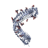 8weeC  8wefC  8wegC  8wehC C: citing same article ( |
|---|---|
| Similar structure data |
-Links
 PDBj
PDBj


- Assembly
| Deposited unit | 
| ||||||||
|---|---|---|---|---|---|---|---|---|---|
| 1 |
| ||||||||
| Unit cell |
|
-Components
-Protein / Non-polymers , 2 types, 213 molecules A
| #1: Protein | Mass: 77498.469 Da / Num. of mol.: 1 / Mutation: N393D Source method: isolated from a genetically manipulated source Source: (gene. exp.)   Spodoptera frugiperda (fall armyworm) / References: UniProt: A0A816I5A6 Spodoptera frugiperda (fall armyworm) / References: UniProt: A0A816I5A6 |
|---|---|
| #7: Water | ChemComp-HOH / Mass: 18.015 Da / Num. of mol.: 212 / Source method: isolated from a natural source / Formula: H2O |
-Sugars , 5 types, 9 molecules 
| #2: Polysaccharide | Source method: isolated from a genetically manipulated source #3: Polysaccharide | beta-D-mannopyranose-(1-4)-2-acetamido-2-deoxy-beta-D-glucopyranose-(1-4)-[alpha-L-fucopyranose-(1- ...beta-D-mannopyranose-(1-4)-2-acetamido-2-deoxy-beta-D-glucopyranose-(1-4)-[alpha-L-fucopyranose-(1-6)]2-acetamido-2-deoxy-beta-D-glucopyranose | Source method: isolated from a genetically manipulated source #4: Polysaccharide | beta-D-mannopyranose-(1-4)-2-acetamido-2-deoxy-beta-D-glucopyranose-(1-4)-2-acetamido-2-deoxy-beta- ...beta-D-mannopyranose-(1-4)-2-acetamido-2-deoxy-beta-D-glucopyranose-(1-4)-2-acetamido-2-deoxy-beta-D-glucopyranose | Source method: isolated from a genetically manipulated source #5: Polysaccharide | 2-acetamido-2-deoxy-beta-D-glucopyranose-(1-4)-[alpha-L-fucopyranose-(1-6)]2-acetamido-2-deoxy-beta- ...2-acetamido-2-deoxy-beta-D-glucopyranose-(1-4)-[alpha-L-fucopyranose-(1-6)]2-acetamido-2-deoxy-beta-D-glucopyranose | Source method: isolated from a genetically manipulated source #6: Sugar |  Type: D-saccharide, beta linking / Mass: 221.208 Da / Num. of mol.: 3 / Source method: obtained synthetically / Formula: C8H15NO6 / Feature type: SUBJECT OF INVESTIGATION Type: D-saccharide, beta linking / Mass: 221.208 Da / Num. of mol.: 3 / Source method: obtained synthetically / Formula: C8H15NO6 / Feature type: SUBJECT OF INVESTIGATION |
|---|
-Details
| Has ligand of interest | Y |
|---|---|
| Has protein modification | Y |
-Experimental details
-Experiment
| Experiment | Method: X-RAY DIFFRACTION / Number of used crystals: 1 |
|---|
- Sample preparation
| Crystal | Density Matthews: 5.73 Å3/Da / Density % sol: 78.54 % |
|---|---|
| Crystal grow | Temperature: 289 K / Method: evaporation / pH: 8.5 Details: 0.2 M Lithium sulfate, 0.1 M Tris pH 8.5, 34% w/v PEG 400 |
-Data collection
| Diffraction | Mean temperature: 80 K / Serial crystal experiment: N |
|---|---|
| Diffraction source | Source: SYNCHROTRON / Site: NFPSS / Beamline: BL19U1 / Wavelength: 0.97853 Å |
| Detector | Type: PSI PILATUS 6M / Detector: PIXEL / Date: Apr 10, 2023 |
| Radiation | Protocol: SINGLE WAVELENGTH / Monochromatic (M) / Laue (L): M / Scattering type: x-ray |
| Radiation wavelength | Wavelength: 0.97853 Å / Relative weight: 1 |
| Reflection | Resolution: 2.7→50 Å / Num. obs: 49088 / % possible obs: 100 % / Redundancy: 5.8 % / CC1/2: 0.98 / Net I/σ(I): 10.7 |
| Reflection shell | Resolution: 2.7→2.8 Å / Num. unique obs: 1468 / CC1/2: 0.57 |
- Processing
| Software |
| |||||||||||||||||||||||||||||||||||||||||||||||||||||||||||||||||||||||||||||||||||||||||||||||||||||||||||||||||||||||
|---|---|---|---|---|---|---|---|---|---|---|---|---|---|---|---|---|---|---|---|---|---|---|---|---|---|---|---|---|---|---|---|---|---|---|---|---|---|---|---|---|---|---|---|---|---|---|---|---|---|---|---|---|---|---|---|---|---|---|---|---|---|---|---|---|---|---|---|---|---|---|---|---|---|---|---|---|---|---|---|---|---|---|---|---|---|---|---|---|---|---|---|---|---|---|---|---|---|---|---|---|---|---|---|---|---|---|---|---|---|---|---|---|---|---|---|---|---|---|---|---|
| Refinement | Method to determine structure: MOLECULAR REPLACEMENT / Resolution: 2.71→36.68 Å / SU ML: 0.32 / Cross valid method: FREE R-VALUE / σ(F): 1.35 / Phase error: 23.02 / Stereochemistry target values: ML
| |||||||||||||||||||||||||||||||||||||||||||||||||||||||||||||||||||||||||||||||||||||||||||||||||||||||||||||||||||||||
| Solvent computation | Shrinkage radii: 0.9 Å / VDW probe radii: 1.1 Å / Solvent model: FLAT BULK SOLVENT MODEL | |||||||||||||||||||||||||||||||||||||||||||||||||||||||||||||||||||||||||||||||||||||||||||||||||||||||||||||||||||||||
| Refinement step | Cycle: LAST / Resolution: 2.71→36.68 Å
| |||||||||||||||||||||||||||||||||||||||||||||||||||||||||||||||||||||||||||||||||||||||||||||||||||||||||||||||||||||||
| Refine LS restraints |
| |||||||||||||||||||||||||||||||||||||||||||||||||||||||||||||||||||||||||||||||||||||||||||||||||||||||||||||||||||||||
| LS refinement shell |
| |||||||||||||||||||||||||||||||||||||||||||||||||||||||||||||||||||||||||||||||||||||||||||||||||||||||||||||||||||||||
| Refinement TLS params. | Method: refined / Refine-ID: X-RAY DIFFRACTION
| |||||||||||||||||||||||||||||||||||||||||||||||||||||||||||||||||||||||||||||||||||||||||||||||||||||||||||||||||||||||
| Refinement TLS group |
|
