Phlebovirus, non structural protein / Rift valley fever virus non structural protein-like / Rift valley fever virus non structural protein (NSs) like / Non-structural protein NS-S
Function and homology information
Biological species
Rift Valley fever virus
Method
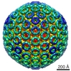
ELECTRON MICROSCOPY / helical reconstruction / cryo EM / Resolution: 2.94 Å
Journal: Cell / Year: 2024 Title: Rift Valley fever virus coordinates the assembly of a programmable E3 ligase to promote viral replication. Authors: Huiling Li / Yulan Zhang / Guibo Rao / Chongtao Zhang / Zhenqiong Guan / Ziyan Huang / Shufen Li / Pierre-Yves Lozach / Sheng Cao / Ke Peng / Abstract: Viruses encode strategies to degrade cellular proteins to promote infection and pathogenesis. Here, we revealed that the non-structural protein NSs of Rift Valley fever virus forms a filamentous E3 ...Viruses encode strategies to degrade cellular proteins to promote infection and pathogenesis. Here, we revealed that the non-structural protein NSs of Rift Valley fever virus forms a filamentous E3 ligase to trigger efficient degradation of targeted proteins. Reconstitution in vitro and cryoelectron microscopy analysis with the 2.9-Å resolution revealed that NSs forms right-handed helical fibrils. The NSs filamentous oligomers associate with the cellular FBXO3 to form a remodeled E3 ligase. The NSs-FBXO3 E3 ligase targets the cellular TFIIH complex through the NSs-P62 interaction, leading to ubiquitination and proteasome-dependent degradation of the TFIIH complex. NSs-FBXO3-triggered TFIIH complex degradation resulted in robust inhibition of antiviral immunity and promoted viral pathogenesis in vivo. Furthermore, it is demonstrated that NSs can be programmed to target additional proteins for proteasome-dependent degradation, serving as a versatile targeted protein degrader. These results showed that a virulence factor forms a filamentous and programmable degradation machinery to induce organized degradation of cellular proteins to promote viral infection.
Component-ID: 1 / Ens-ID: ens_1 / Beg auth comp-ID: ASP / Beg label comp-ID: ASP / End auth comp-ID: PRO / End label comp-ID: PRO / Auth seq-ID: 2 - 244 / Label seq-ID: 11 - 253
Dom-ID
Auth asym-ID
Label asym-ID
d_1
B
B
d_2
A
A
-
Components
#1: Protein
Non-structuralproteinNS-S
Mass: 30870.221 Da / Num. of mol.: 2 Source method: isolated from a genetically manipulated source Source: (gene. exp.) Rift Valley fever virus / Gene: NSs / Production host: Escherichia coli BL21(DE3) (bacteria) / Strain (production host): BL21(DE3) / References: UniProt: A2SZZ6
Has protein modification
N
-
Experimental details
-
Experiment
Experiment
Method: ELECTRON MICROSCOPY
EM experiment
Aggregation state: HELICAL ARRAY / 3D reconstruction method: helical reconstruction
-
Sample preparation
Component
Name: The self-assembly of RVFV NSs protein / Type: COMPLEX / Entity ID: all / Source: RECOMBINANT
Molecular weight
Experimental value: NO
Source (natural)
Organism: Rift Valley fever virus
Source (recombinant)
Organism: Escherichia coli BL21(DE3) (bacteria)
Buffer solution
pH: 7.4
Specimen
Embedding applied: NO / Shadowing applied: NO / Staining applied: NO / Vitrification applied: YES
Vitrification
Cryogen name: ETHANE
-
Electron microscopy imaging
Microscopy
Model: JEOL CRYO ARM 300
Electron gun
Electron source: FIELD EMISSION GUN / Accelerating voltage: 300 kV / Illumination mode: FLOOD BEAM
In the structure databanks used in Yorodumi, some data are registered as the other names, "COVID-19 virus" and "2019-nCoV". Here are the details of the virus and the list of structure data.
Jan 31, 2019. EMDB accession codes are about to change! (news from PDBe EMDB page)
EMDB accession codes are about to change! (news from PDBe EMDB page)
The allocation of 4 digits for EMDB accession codes will soon come to an end. Whilst these codes will remain in use, new EMDB accession codes will include an additional digit and will expand incrementally as the available range of codes is exhausted. The current 4-digit format prefixed with “EMD-” (i.e. EMD-XXXX) will advance to a 5-digit format (i.e. EMD-XXXXX), and so on. It is currently estimated that the 4-digit codes will be depleted around Spring 2019, at which point the 5-digit format will come into force.
The EM Navigator/Yorodumi systems omit the EMD- prefix.
Related info.:Q: What is EMD? / ID/Accession-code notation in Yorodumi/EM Navigator
Yorodumi is a browser for structure data from EMDB, PDB, SASBDB, etc.
This page is also the successor to EM Navigator detail page, and also detail information page/front-end page for Omokage search.
The word "yorodu" (or yorozu) is an old Japanese word meaning "ten thousand". "mi" (miru) is to see.
Related info.:EMDB / PDB / SASBDB / Comparison of 3 databanks / Yorodumi Search / Aug 31, 2016. New EM Navigator & Yorodumi / Yorodumi Papers / Jmol/JSmol / Function and homology information / Changes in new EM Navigator and Yorodumi
 Movie
Movie Controller


 Open data
 Basic information
 Components
 Keywords
 Function and homology information
 Rift Valley fever virus
 Authors
 China, 1items
  Citation
 Structure visualization
 Downloads & links
 Other downloads
 PDBj
PDBj Assembly

 Sample preparation
 Electron microscopy imaging
 FIELD EMISSION GUN / Accelerating voltage: 300 kV / Illumination mode: FLOOD BEAM
 Processing