 Movie
Movie Controller


+ Open data

- Basic information
| Entry |  | |||||||||
|---|---|---|---|---|---|---|---|---|---|---|
| Title | NSs filament formation determines RVFV pathogenesis | |||||||||
 Map data Map data | EM map | |||||||||
 Sample Sample |
| |||||||||
 Keywords Keywords | Self-assembly / helical structure / VIRAL PROTEIN | |||||||||
| Function / homology | Phlebovirus, non structural protein / Rift valley fever virus non structural protein-like / Rift valley fever virus non structural protein (NSs) like / Non-structural protein NS-S Function and homology information Function and homology information | |||||||||
| Biological species |  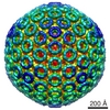 Rift Valley fever virus Rift Valley fever virus | |||||||||
| Method | helical reconstruction / cryo EM / Resolution: 2.94 Å | |||||||||
 Authors Authors | Li H / Rao G / Cao S / Peng K | |||||||||
| Funding support |  China, 1 items China, 1 items
| |||||||||
 Citation Citation | Journal: Cell / Year: 2024 Title: Rift Valley fever virus coordinates the assembly of a programmable E3 ligase to promote viral replication. Authors: Huiling Li / Yulan Zhang / Guibo Rao / Chongtao Zhang / Zhenqiong Guan / Ziyan Huang / Shufen Li / Pierre-Yves Lozach / Sheng Cao / Ke Peng /  Abstract: Viruses encode strategies to degrade cellular proteins to promote infection and pathogenesis. Here, we revealed that the non-structural protein NSs of Rift Valley fever virus forms a filamentous E3 ...Viruses encode strategies to degrade cellular proteins to promote infection and pathogenesis. Here, we revealed that the non-structural protein NSs of Rift Valley fever virus forms a filamentous E3 ligase to trigger efficient degradation of targeted proteins. Reconstitution in vitro and cryoelectron microscopy analysis with the 2.9-Å resolution revealed that NSs forms right-handed helical fibrils. The NSs filamentous oligomers associate with the cellular FBXO3 to form a remodeled E3 ligase. The NSs-FBXO3 E3 ligase targets the cellular TFIIH complex through the NSs-P62 interaction, leading to ubiquitination and proteasome-dependent degradation of the TFIIH complex. NSs-FBXO3-triggered TFIIH complex degradation resulted in robust inhibition of antiviral immunity and promoted viral pathogenesis in vivo. Furthermore, it is demonstrated that NSs can be programmed to target additional proteins for proteasome-dependent degradation, serving as a versatile targeted protein degrader. These results showed that a virulence factor forms a filamentous and programmable degradation machinery to induce organized degradation of cellular proteins to promote viral infection. | |||||||||
| History |
|
- Structure visualization
| Supplemental images |
|---|

- Downloads & links
-EMDB archive
| Map data | emd_37443.map.gz | 16.7 MB | EMDB map data format | |
|---|---|---|---|---|
| Header (meta data) | emd-37443-v30.xmlemd-37443.xml | 13.4 KB 13.4 KB | Display Display | EMDB header |
| FSC (resolution estimation) | emd_37443_fsc.xml | 8.4 KB | Display | FSC data file |
| Images |  emd_37443.png emd_37443.png | 127.1 KB | ||
| Filedesc metadata | emd-37443.cif.gz | 5.2 KB | ||
| Others | emd_37443_half_map_1.map.gzemd_37443_half_map_2.map.gz | 59.4 MB 59.4 MB | ||
| Archive directory |  http://ftp.pdbj.org/pub/emdb/structures/EMD-37443ftp://ftp.pdbj.org/pub/emdb/structures/EMD-37443 http://ftp.pdbj.org/pub/emdb/structures/EMD-37443ftp://ftp.pdbj.org/pub/emdb/structures/EMD-37443 | HTTPS FTP |
-Related structure data
| Related structure data |  8wcmMC M: atomic model generated by this map C: citing same article ( |
|---|---|
| Similar structure data |
-Links
| EMDB pages | EMDB (EBI/PDBe) / EMDataResource |
|---|
-Map
| File | Download / File: emd_37443.map.gz / Format: CCP4 / Size: 64 MB / Type: IMAGE STORED AS FLOATING POINT NUMBER (4 BYTES) | ||||||||||||||||||||||||||||||||||||
|---|---|---|---|---|---|---|---|---|---|---|---|---|---|---|---|---|---|---|---|---|---|---|---|---|---|---|---|---|---|---|---|---|---|---|---|---|---|
| Annotation | EM map | ||||||||||||||||||||||||||||||||||||
| Projections & slices | Image control
Images are generated by Spider. | ||||||||||||||||||||||||||||||||||||
| Voxel size | X=Y=Z: 0.95 Å | ||||||||||||||||||||||||||||||||||||
| Density |
| ||||||||||||||||||||||||||||||||||||
| Symmetry | Space group: 1 | ||||||||||||||||||||||||||||||||||||
| Details | EMDB XML:
|
 Z (Sec.)
 Y (Row.)
 X (Col.)


-Supplemental data
-Half map: EM half map
| File | emd_37443_half_map_1.map | ||||||||||||
|---|---|---|---|---|---|---|---|---|---|---|---|---|---|
| Annotation | EM half map | ||||||||||||
| Projections & Slices |
| ||||||||||||
| Density Histograms |


-Half map: EM half map
| File | emd_37443_half_map_2.map | ||||||||||||
|---|---|---|---|---|---|---|---|---|---|---|---|---|---|
| Annotation | EM half map | ||||||||||||
| Projections & Slices |
| ||||||||||||
| Density Histograms |


- Sample components
-Entire : The self-assembly of RVFV NSs protein
| Entire | Name: The self-assembly of RVFV NSs protein |
|---|---|
| Components |
|
-Supramolecule #1: The self-assembly of RVFV NSs protein
| Supramolecule | Name: The self-assembly of RVFV NSs protein / type: complex / ID: 1 / Parent: 0 / Macromolecule list: all |
|---|---|
| Source (natural) | Organism: Rift Valley fever virus |
-Macromolecule #1: Non-structural protein NS-S
| Macromolecule | Name: Non-structural protein NS-S / type: protein_or_peptide / ID: 1 / Number of copies: 2 / Enantiomer: LEVO |
|---|---|
| Source (natural) | Organism: Rift Valley fever virus |
| Molecular weight | Theoretical: 30.870221 KDa |
| Recombinant expression | Organism:  |
| Sequence | String: HHHHHHGSGG DYFPVISVDL QSGRRVVSVE YIRGDGPPRI PYSMVGPCCV FLMHHRPSHE VRLRFSDFYN VGEFPYRVGL GDFASNVAP PPAKPFQRLI DLIGHMTLSD FTRFPNLKEA ISWPLGEPSL AFFDLSSTRV HRNDDIRRDQ IATLAMRSCK I TNDLEDSF ...String: HHHHHHGSGG DYFPVISVDL QSGRRVVSVE YIRGDGPPRI PYSMVGPCCV FLMHHRPSHE VRLRFSDFYN VGEFPYRVGL GDFASNVAP PPAKPFQRLI DLIGHMTLSD FTRFPNLKEA ISWPLGEPSL AFFDLSSTRV HRNDDIRRDQ IATLAMRSCK I TNDLEDSF VGLHRMIVTE AILRGIDLCL LPGFDLMYEV AHVQCVRLLQ AAKEDISNAV VPNSALIALM EESLMLRSSL PS MMGRNNW IPVVPPIPDV EMESEEESDD DGFVEVD UniProtKB: Non-structural protein NS-S |
-Experimental details
-Structure determination
| Method | cryo EM |
|---|---|
 Processing Processing | helical reconstruction |
| Aggregation state | helical array |
-Sample preparation
| Buffer | pH: 7.4 |
|---|---|
| Vitrification | Cryogen name: ETHANE |
- Electron microscopy
| Microscope | JEOL CRYO ARM 300 |
|---|---|
| Image recording | Film or detector model: GATAN K3 (6k x 4k) / Average electron dose: 40.0 e/Å2 |
| Electron beam | Acceleration voltage: 300 kV / Electron source:  FIELD EMISSION GUN FIELD EMISSION GUN |
| Electron optics | Illumination mode: FLOOD BEAM / Imaging mode: BRIGHT FIELD / Nominal defocus max: 2.5 µm / Nominal defocus min: 0.5 µm |
-Image processing
| Final reconstruction | Applied symmetry - Helical parameters - Δz: 14.584 Å Applied symmetry - Helical parameters - Δ&Phi: 83.437 ° Applied symmetry - Helical parameters - Axial symmetry: C1 (asymmetric) Resolution.type: BY AUTHOR / Resolution: 2.94 Å / Resolution method: FSC 0.143 CUT-OFF / Number images used: 617585 |
|---|---|
| Startup model | Type of model: NONE |
| Final angle assignment | Type: NOT APPLICABLE |
| FSC plot (resolution estimation) |  |
