 Movie
Movie Controller

[English] 日本語
 Yorodumi
- PDB-8w72: Crystal structure of a P450 enzyme DmlH that catalyze intramolecu... -
+ Open data

- Basic information
| Entry | Database: PDB / ID: 8w72 | ||||||||||||
|---|---|---|---|---|---|---|---|---|---|---|---|---|---|
| Title | Crystal structure of a P450 enzyme DmlH that catalyze intramolecular phenol coupling in the biosynthesis of cihanmycins | ||||||||||||
 Components Components | Cytochrome P450 | ||||||||||||
 Keywords Keywords | BIOSYNTHETIC PROTEIN / P450 enzyme / intramolecular phenol coupling / cihanmycins | ||||||||||||
| Function / homology | PROTOPORPHYRIN IX CONTAINING FE Function and homology information Function and homology information | ||||||||||||
| Biological species |  Streptomyces sp. DM14 (bacteria) Streptomyces sp. DM14 (bacteria) | ||||||||||||
| Method |  X-RAY DIFFRACTION / SYNCHROTRON / MOLECULAR REPLACEMENT / Resolution: 3.25 Å X-RAY DIFFRACTION / SYNCHROTRON / MOLECULAR REPLACEMENT / Resolution: 3.25 Å | ||||||||||||
 Authors Authors | Fang, C. / Zhang, L. / Zhu, Y. / Zhang, C. | ||||||||||||
| Funding support |  China, 3items China, 3items
| ||||||||||||
 Citation Citation | Journal: J.Am.Chem.Soc. / Year: 2024 Title: Discovery and Biosynthesis of Cihanmycins Reveal Cytochrome P450-Catalyzed Intramolecular C-O Phenol Coupling Reactions. Authors: Fang, C. / Zhang, L. / Wang, Y. / Xiong, W. / Yan, Z. / Zhang, W. / Zhang, Q. / Wang, B. / Zhu, Y. / Zhang, C. | ||||||||||||
| History |
|
- Structure visualization
| Structure viewer | Molecule: MolmilJmol/JSmol |
|---|
- Downloads & links
-Download
| PDBx/mmCIF format | 8w72.cif.gz | 91.4 KB | Display | PDBx/mmCIF format |
|---|---|---|---|---|
| PDB format | pdb8w72.ent.gz | 66.1 KB | Display | PDB format |
| PDBx/mmJSON format | 8w72.json.gz | Tree view | PDBx/mmJSON format | |
| Others |  Other downloads Other downloads |
-Validation report
| Arichive directory | https://data.pdbj.org/pub/pdb/validation_reports/w7/8w72ftp://data.pdbj.org/pub/pdb/validation_reports/w7/8w72 | HTTPS FTP |
|---|
-Related structure data

-Links
 PDBj
PDBj- Assembly
| Deposited unit | 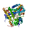
| ||||||||
|---|---|---|---|---|---|---|---|---|---|
| 1 |
| ||||||||
| Unit cell |
|
-Components
| #1: Protein | Mass: 46858.555 Da / Num. of mol.: 1 / Source method: isolated from a natural source / Source: (natural) Streptomyces sp. DM14 (bacteria) |
|---|---|
| #2: Chemical | ChemComp-HEM /   Mass: 616.487 Da / Num. of mol.: 1 / Source method: obtained synthetically / Formula: C34H32FeN4O4 / Feature type: SUBJECT OF INVESTIGATION Mass: 616.487 Da / Num. of mol.: 1 / Source method: obtained synthetically / Formula: C34H32FeN4O4 / Feature type: SUBJECT OF INVESTIGATION |
| Has ligand of interest | Y |
| Has protein modification | N |
-Experimental details
-Experiment
| Experiment | Method: X-RAY DIFFRACTION / Number of used crystals: 1 |
|---|
- Sample preparation
| Crystal | Density Matthews: 2.08 Å3/Da / Density % sol: 40.94 % |
|---|---|
| Crystal grow | Temperature: 289 K / Method: vapor diffusion, sitting drop Details: 0.3 M CaCl2, 0.3 M MgCl2, 0.5 M MES, 0.5 M imidazole, 37.5% w/v PEG 1K_PEG 3350_MPD, pH 6.5 |
-Data collection
| Diffraction | Mean temperature: 100 K / Serial crystal experiment: N |
|---|---|
| Diffraction source | Source: SYNCHROTRON / Site: SSRF / Beamline: BL02U1 / Wavelength: 0.979183 Å |
| Detector | Type: DECTRIS EIGER2 S 9M / Detector: PIXEL / Date: May 21, 2023 |
| Radiation | Protocol: SINGLE WAVELENGTH / Monochromatic (M) / Laue (L): M / Scattering type: x-ray |
| Radiation wavelength | Wavelength: 0.979183 Å / Relative weight: 1 |
| Reflection | Resolution: 3.25→53.49 Å / Num. obs: 6132 / % possible obs: 92.4 % / Redundancy: 7.5 % / CC1/2: 0.994 / Rmerge(I) obs: 0.15 / Rpim(I) all: 0.058 / Rrim(I) all: 0.162 / Χ2: 0.93 / Net I/σ(I): 9.5 / Num. measured all: 45745 |
| Reflection shell | Resolution: 3.25→3.43 Å / % possible obs: 91.3 % / Redundancy: 4.4 % / Rmerge(I) obs: 0.541 / Num. measured all: 3695 / Num. unique obs: 844 / CC1/2: 0.805 / Rpim(I) all: 0.26 / Rrim(I) all: 0.605 / Χ2: 0.75 / Net I/σ(I) obs: 2.3 |
- Processing
| Software |
| ||||||||||||||||||||||||
|---|---|---|---|---|---|---|---|---|---|---|---|---|---|---|---|---|---|---|---|---|---|---|---|---|---|
| Refinement | Method to determine structure: MOLECULAR REPLACEMENT / Resolution: 3.25→50.22 Å / SU ML: 0.42 / Cross valid method: FREE R-VALUE / σ(F): 1.36 / Phase error: 17.32 / Stereochemistry target values: ML
| ||||||||||||||||||||||||
| Solvent computation | Shrinkage radii: 0.9 Å / VDW probe radii: 1.11 Å / Solvent model: FLAT BULK SOLVENT MODEL | ||||||||||||||||||||||||
| Refinement step | Cycle: LAST / Resolution: 3.25→50.22 Å
| ||||||||||||||||||||||||
| Refine LS restraints |
| ||||||||||||||||||||||||
| LS refinement shell |
|
