 Movie
Movie Controller

[English] 日本語
 Yorodumi
- PDB-8w6z: Substrate-bound crystal structure of a P450 enzyme DmlH that cata... -
+ Open data

- Basic information
| Entry | Database: PDB / ID: 8w6z | ||||||||||||
|---|---|---|---|---|---|---|---|---|---|---|---|---|---|
| Title | Substrate-bound crystal structure of a P450 enzyme DmlH that catalyze intramolecular phenol coupling in the biosynthesis of cihanmycins | ||||||||||||
 Components Components |
| ||||||||||||
 Keywords Keywords | BIOSYNTHETIC PROTEIN / P450 enzyme / intramolecular phenol coupling / cihanmycins | ||||||||||||
| Function / homology | : / PROTOPORPHYRIN IX CONTAINING FE / :  Function and homology information Function and homology information | ||||||||||||
| Biological species |  Streptomyces sp. DM14 (bacteria) Streptomyces sp. DM14 (bacteria) | ||||||||||||
| Method |  X-RAY DIFFRACTION / MOLECULAR REPLACEMENT / Resolution: 2.2 Å X-RAY DIFFRACTION / MOLECULAR REPLACEMENT / Resolution: 2.2 Å | ||||||||||||
 Authors Authors | Fang, C. / Zhang, L. / Zhu, Y. / Zhang, C. | ||||||||||||
| Funding support |  China, 3items China, 3items
| ||||||||||||
 Citation Citation | Journal: J.Am.Chem.Soc. / Year: 2024 Title: Discovery and Biosynthesis of Cihanmycins Reveal Cytochrome P450-Catalyzed Intramolecular C-O Phenol Coupling Reactions. Authors: Fang, C. / Zhang, L. / Wang, Y. / Xiong, W. / Yan, Z. / Zhang, W. / Zhang, Q. / Wang, B. / Zhu, Y. / Zhang, C. | ||||||||||||
| History |
|
- Structure visualization
| Structure viewer | Molecule: MolmilJmol/JSmol |
|---|
- Downloads & links
-Download
| PDBx/mmCIF format | 8w6z.cif.gz | 107.1 KB | Display | PDBx/mmCIF format |
|---|---|---|---|---|
| PDB format | pdb8w6z.ent.gz | Display | PDB format | |
| PDBx/mmJSON format | 8w6z.json.gz | Tree view | PDBx/mmJSON format | |
| Others |  Other downloads Other downloads |
-Validation report
| Arichive directory | https://data.pdbj.org/pub/pdb/validation_reports/w6/8w6zftp://data.pdbj.org/pub/pdb/validation_reports/w6/8w6z | HTTPS FTP |
|---|
-Related structure data

-Links
 PDBj
PDBj- Assembly
| Deposited unit | 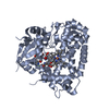
| ||||||||
|---|---|---|---|---|---|---|---|---|---|
| 1 |
| ||||||||
| Unit cell |
|
-Components
| #1: Protein | Mass: 46858.555 Da / Num. of mol.: 1 / Source method: isolated from a natural source / Source: (natural) Streptomyces sp. DM14 (bacteria) |
|---|---|
| #2: Protein/peptide | Type: Cyclic peptide / Class: Unknown / Mass: 1227.275 Da / Num. of mol.: 1 / Source method: isolated from a natural source / Source: (natural) Streptomyces sp. DM14 (bacteria) / References: BIRD: PRD_002429 |
| #3: Chemical | ChemComp-HEM /   Mass: 616.487 Da / Num. of mol.: 1 / Source method: obtained synthetically / Formula: C34H32FeN4O4 Mass: 616.487 Da / Num. of mol.: 1 / Source method: obtained synthetically / Formula: C34H32FeN4O4 |
| #4: Chemical | ChemComp-R8L / ( Type: Cyclic peptide / Class: Unknown / Mass: 291.299 Da / Num. of mol.: 1 / Source method: obtained synthetically / Formula: C15H17NO5 / Feature type: SUBJECT OF INVESTIGATION / References: BIRD: PRD_002429 |
| #5: Water | ChemComp-HOH /  Mass: 18.015 Da / Num. of mol.: 193 / Source method: isolated from a natural source / Formula: H2O Mass: 18.015 Da / Num. of mol.: 193 / Source method: isolated from a natural source / Formula: H2O |
| Compound details | Author stated that they first isolated this compound from the Streptomyces lividans SBT18 host ...Author stated that they first isolated this compound from the Streptomyces lividans SBT18 host haboring the biosynthetic gene cluster of cihanmycin (GenBank accession no.TQJ01344-TQJ01379), with the cytochrome p450 monooxygenase gene cih33 inactivated. |
| Has ligand of interest | Y |
| Has protein modification | Y |
-Experimental details
-Experiment
| Experiment | Method: X-RAY DIFFRACTION / Number of used crystals: 1 |
|---|
- Sample preparation
| Crystal | Density Matthews: 1.99 Å3/Da / Density % sol: 38.15 % |
|---|---|
| Crystal grow | Temperature: 289 K / Method: vapor diffusion, sitting drop / pH: 6.5 Details: 3 M CaCl2, 0.3 M MgCl2, 0.5 M MES, 0.5 M imidazole, 37.5% w/v PEG 1K_PEG 3350_MPD, pH 6.5 |
-Data collection
| Diffraction | Mean temperature: 100 K / Serial crystal experiment: N |
|---|---|
| Diffraction source | Source: ROTATING ANODE / Type: RIGAKU / Wavelength: 1.5418 Å |
| Detector | Type: DECTRIS PILATUS 200K / Detector: PIXEL / Date: Jun 19, 2023 |
| Radiation | Protocol: SINGLE WAVELENGTH / Monochromatic (M) / Laue (L): M / Scattering type: x-ray |
| Radiation wavelength | Wavelength: 1.5418 Å / Relative weight: 1 |
| Reflection | Resolution: 2.2→19.68 Å / Num. obs: 20263 / % possible obs: 99.9 % / Redundancy: 9.3 % / CC1/2: 0.998 / Rmerge(I) obs: 0.095 / Rpim(I) all: 0.032 / Rrim(I) all: 0.101 / Net I/σ(I): 15.8 / Num. measured all: 188014 |
| Reflection shell | Resolution: 2.2→2.32 Å / % possible obs: 100 % / Redundancy: 5 % / Rmerge(I) obs: 0.49 / Num. measured all: 14569 / Num. unique obs: 2903 / CC1/2: 0.828 / Rpim(I) all: 0.24 / Rrim(I) all: 0.548 / Net I/σ(I) obs: 3.1 |
- Processing
| Software |
| ||||||||||||||||||||||||||||||||||||||||||||||||||||||||
|---|---|---|---|---|---|---|---|---|---|---|---|---|---|---|---|---|---|---|---|---|---|---|---|---|---|---|---|---|---|---|---|---|---|---|---|---|---|---|---|---|---|---|---|---|---|---|---|---|---|---|---|---|---|---|---|---|---|
| Refinement | Method to determine structure: MOLECULAR REPLACEMENT / Resolution: 2.2→19.68 Å / SU ML: 0.17 / Cross valid method: FREE R-VALUE / σ(F): 1.34 / Phase error: 20.63 / Stereochemistry target values: ML
| ||||||||||||||||||||||||||||||||||||||||||||||||||||||||
| Solvent computation | Shrinkage radii: 0.9 Å / VDW probe radii: 1.11 Å / Solvent model: FLAT BULK SOLVENT MODEL | ||||||||||||||||||||||||||||||||||||||||||||||||||||||||
| Refinement step | Cycle: LAST / Resolution: 2.2→19.68 Å
| ||||||||||||||||||||||||||||||||||||||||||||||||||||||||
| Refine LS restraints |
| ||||||||||||||||||||||||||||||||||||||||||||||||||||||||
| LS refinement shell |
|
