 Movie
Movie Controller


+ Open data

- Basic information
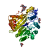
| Entry | Database: PDB / ID: 8w6p | ||||||
|---|---|---|---|---|---|---|---|
| Title | Crystal structure of dimeric murine SMPDL3A | ||||||
 Components Components | Acid sphingomyelinase-like phosphodiesterase 3a | ||||||
 Keywords Keywords | HYDROLASE / innate immunity / lipid metabolism | ||||||
| Function / homology |  Function and homology information Function and homology informationnucleoside triphosphate catabolic process / Hydrolases; Acting on ester bonds; Phosphoric-diester hydrolases / phosphoric diester hydrolase activity / negative regulation of cGAS/STING signaling pathway / nucleoside-triphosphate phosphatase / ATP hydrolysis activity / : / extracellular region / zinc ion binding Similarity search - Function | ||||||
| Biological species |  | ||||||
| Method |  X-RAY DIFFRACTION / MOLECULAR REPLACEMENT / Resolution: 1.91 Å X-RAY DIFFRACTION / MOLECULAR REPLACEMENT / Resolution: 1.91 Å | ||||||
 Authors Authors | Zhang, C. / Liu, P. / Fan, S. / Hou, Y. | ||||||
| Funding support |  China, 1items China, 1items
| ||||||
 Citation Citation | Journal: Immunity / Year: 2023 Title: SMPDL3A is a cGAMP-degrading enzyme induced by LXR-mediated lipid metabolism to restrict cGAS-STING DNA sensing. Authors: Hou, Y. / Wang, Z. / Liu, P. / Wei, X. / Zhang, Z. / Fan, S. / Zhang, L. / Han, F. / Song, Y. / Chu, L. / Zhang, C. | ||||||
| History |
|
- Structure visualization
| Structure viewer | Molecule: MolmilJmol/JSmol |
|---|
- Downloads & links
-Download
| PDBx/mmCIF format | 8w6p.cif.gz | 206.6 KB | Display | PDBx/mmCIF format |
|---|---|---|---|---|
| PDB format | pdb8w6p.ent.gz | 158.9 KB | Display | PDB format |
| PDBx/mmJSON format | 8w6p.json.gz | Tree view | PDBx/mmJSON format | |
| Others |  Other downloads Other downloads |
-Validation report
| Arichive directory | https://data.pdbj.org/pub/pdb/validation_reports/w6/8w6pftp://data.pdbj.org/pub/pdb/validation_reports/w6/8w6p | HTTPS FTP |
|---|
-Related structure data
-Links
 PDBj
PDBj

- Assembly
| Deposited unit | 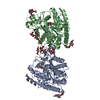
| ||||||||
|---|---|---|---|---|---|---|---|---|---|
| 1 |
| ||||||||
| Unit cell |
|
-Components
-Protein , 1 types, 2 molecules AB
| #1: Protein | Mass: 46664.617 Da / Num. of mol.: 2 Source method: isolated from a genetically manipulated source Source: (gene. exp.)   Spodoptera frugiperda (fall armyworm) Spodoptera frugiperda (fall armyworm)References: UniProt: P70158, Hydrolases; Acting on ester bonds; Phosphoric-diester hydrolases |
|---|
-Sugars , 5 types, 11 molecules 

| #2: Polysaccharide | Source method: isolated from a genetically manipulated source #3: Polysaccharide | alpha-L-fucopyranose-(1-6)-2-acetamido-2-deoxy-beta-D-glucopyranose | #4: Polysaccharide | beta-D-mannopyranose-(1-4)-2-acetamido-2-deoxy-beta-D-glucopyranose-(1-4)-2-acetamido-2-deoxy-beta- ...beta-D-mannopyranose-(1-4)-2-acetamido-2-deoxy-beta-D-glucopyranose-(1-4)-2-acetamido-2-deoxy-beta-D-glucopyranose | #6: Sugar | ChemComp-NAG /  Type: D-saccharide, beta linking / Mass: 221.208 Da / Num. of mol.: 6 / Source method: obtained synthetically / Formula: C8H15NO6 / Feature type: SUBJECT OF INVESTIGATION Type: D-saccharide, beta linking / Mass: 221.208 Da / Num. of mol.: 6 / Source method: obtained synthetically / Formula: C8H15NO6 / Feature type: SUBJECT OF INVESTIGATION#8: Sugar | ChemComp-FUC / |  Type: L-saccharide, alpha linking / Mass: 164.156 Da / Num. of mol.: 1 / Source method: obtained synthetically / Formula: C6H12O5 / Feature type: SUBJECT OF INVESTIGATION Type: L-saccharide, alpha linking / Mass: 164.156 Da / Num. of mol.: 1 / Source method: obtained synthetically / Formula: C6H12O5 / Feature type: SUBJECT OF INVESTIGATION |
|---|
-Non-polymers , 4 types, 810 molecules 


| #5: Chemical | ChemComp-YA4 / [( Type: RNA linking / Mass: 230.176 Da / Num. of mol.: 1 / Source method: obtained synthetically / Formula: C5H11O6PS / Feature type: SUBJECT OF INVESTIGATION Type: RNA linking / Mass: 230.176 Da / Num. of mol.: 1 / Source method: obtained synthetically / Formula: C5H11O6PS / Feature type: SUBJECT OF INVESTIGATION | ||||
|---|---|---|---|---|---|
| #7: Chemical | ChemComp-ZN /  Mass: 65.409 Da / Num. of mol.: 4 / Source method: obtained synthetically / Formula: Zn / Feature type: SUBJECT OF INVESTIGATION Mass: 65.409 Da / Num. of mol.: 4 / Source method: obtained synthetically / Formula: Zn / Feature type: SUBJECT OF INVESTIGATION#9: Chemical | ChemComp-PO4 / |  Mass: 94.971 Da / Num. of mol.: 1 / Source method: obtained synthetically / Formula: PO4 / Feature type: SUBJECT OF INVESTIGATION Mass: 94.971 Da / Num. of mol.: 1 / Source method: obtained synthetically / Formula: PO4 / Feature type: SUBJECT OF INVESTIGATION#10: Water | ChemComp-HOH / | Mass: 18.015 Da / Num. of mol.: 804 / Source method: isolated from a natural source / Formula: H2O |
-Details
| Has ligand of interest | Y |
|---|---|
| Has protein modification | Y |
-Experimental details
-Experiment
| Experiment | Method: X-RAY DIFFRACTION / Number of used crystals: 1 |
|---|
- Sample preparation
| Crystal | Density Matthews: 3.08 Å3/Da / Density % sol: 60.1 % |
|---|---|
| Crystal grow | Temperature: 291 K / Method: vapor diffusion Details: 0.1 M sodium citrate [pH 5.5], 0.2M ammonium sulfate and 30% PEG 4000 |
-Data collection
| Diffraction | Mean temperature: 100 K / Serial crystal experiment: N |
|---|---|
| Diffraction source | Source: ROTATING ANODE / Type: RIGAKU / Wavelength: 1.5418 Å |
| Detector | Type: RIGAKU HyPix-6000HE / Detector: PIXEL / Date: Mar 16, 2022 |
| Radiation | Protocol: SINGLE WAVELENGTH / Monochromatic (M) / Laue (L): M / Scattering type: x-ray |
| Radiation wavelength | Wavelength: 1.5418 Å / Relative weight: 1 |
| Reflection | Resolution: 1.91→20.27 Å / Num. obs: 89995 / % possible obs: 98.6 % / Redundancy: 7.5 % / Biso Wilson estimate: 27 Å2 / CC1/2: 0.999 / Rmerge(I) obs: 0.101 / Net I/σ(I): 17.9 |
| Reflection shell | Resolution: 1.91→1.94 Å / Redundancy: 3.5 % / Rmerge(I) obs: 0.768 / Mean I/σ(I) obs: 1 / Num. unique obs: 3909 / CC1/2: 0.597 / % possible all: 85.1 |
- Processing
| Software |
| |||||||||||||||||||||||||||||||||||||||||||||||||||||||||||||||||||||||||||||||||||||||||||||||||||||||||
|---|---|---|---|---|---|---|---|---|---|---|---|---|---|---|---|---|---|---|---|---|---|---|---|---|---|---|---|---|---|---|---|---|---|---|---|---|---|---|---|---|---|---|---|---|---|---|---|---|---|---|---|---|---|---|---|---|---|---|---|---|---|---|---|---|---|---|---|---|---|---|---|---|---|---|---|---|---|---|---|---|---|---|---|---|---|---|---|---|---|---|---|---|---|---|---|---|---|---|---|---|---|---|---|---|---|---|
| Refinement | Method to determine structure: MOLECULAR REPLACEMENT / Resolution: 1.91→20.27 Å / SU ML: 0.27 / Cross valid method: FREE R-VALUE / σ(F): 1.34 / Phase error: 28.05 / Stereochemistry target values: ML
| |||||||||||||||||||||||||||||||||||||||||||||||||||||||||||||||||||||||||||||||||||||||||||||||||||||||||
| Solvent computation | Shrinkage radii: 0.9 Å / VDW probe radii: 1.11 Å / Solvent model: FLAT BULK SOLVENT MODEL | |||||||||||||||||||||||||||||||||||||||||||||||||||||||||||||||||||||||||||||||||||||||||||||||||||||||||
| Refinement step | Cycle: LAST / Resolution: 1.91→20.27 Å
| |||||||||||||||||||||||||||||||||||||||||||||||||||||||||||||||||||||||||||||||||||||||||||||||||||||||||
| Refine LS restraints |
| |||||||||||||||||||||||||||||||||||||||||||||||||||||||||||||||||||||||||||||||||||||||||||||||||||||||||
| LS refinement shell |
|
