 Movie
Movie Controller

[English] 日本語
 Yorodumi
- PDB-8vti: Latrophilin-3 (ADGRL3) HormR and GAIN domains in the context of t... -
+ Open data

- Basic information
| Entry | Database: PDB / ID: 8vti | ||||||||||||||||||||||||
|---|---|---|---|---|---|---|---|---|---|---|---|---|---|---|---|---|---|---|---|---|---|---|---|---|---|
| Title | Latrophilin-3 (ADGRL3) HormR and GAIN domains in the context of the holoreceptor | ||||||||||||||||||||||||
 Components Components |
| ||||||||||||||||||||||||
 Keywords Keywords | SIGNALING PROTEIN / adhesion GPCR / GAIN domain / cell-cell adhesion / synapse | ||||||||||||||||||||||||
| Function / homology |  Function and homology information Function and homology informationRho-activating G protein-coupled receptor signaling pathway / excitatory synapse assembly / cell adhesion mediator activity / : / synapse assembly / molecular condensate scaffold activity / neuron migration / G protein-coupled receptor activity / cell-cell junction / carbohydrate binding ...Rho-activating G protein-coupled receptor signaling pathway / excitatory synapse assembly / cell adhesion mediator activity / : / synapse assembly / molecular condensate scaffold activity / neuron migration / G protein-coupled receptor activity / cell-cell junction / carbohydrate binding / adenylate cyclase-activating G protein-coupled receptor signaling pathway / postsynaptic membrane / cell surface receptor signaling pathway / G protein-coupled receptor signaling pathway / axon / calcium ion binding / glutamatergic synapse / membrane / plasma membrane Similarity search - Function | ||||||||||||||||||||||||
| Biological species |  Homo sapiens (human) Homo sapiens (human)synthetic construct (others) | ||||||||||||||||||||||||
| Method | ELECTRON MICROSCOPY / single particle reconstruction / cryo EM / Resolution: 3.9 Å | ||||||||||||||||||||||||
 Authors Authors | Kordon, S.P. / Bandekar, S.J. / Arac, D. | ||||||||||||||||||||||||
| Funding support |  United States, 3items United States, 3items
| ||||||||||||||||||||||||
 Citation Citation | Journal: Nat Commun / Year: 2024 Title: Conformational coupling between extracellular and transmembrane domains modulates holo-adhesion GPCR function. Authors: Szymon P Kordon / Kristina Cechova / Sumit J Bandekar / Katherine Leon / Przemysław Dutka / Gracie Siffer / Anthony A Kossiakoff / Reza Vafabakhsh / Demet Araç / Abstract: Adhesion G Protein-Coupled Receptors (aGPCRs) are key cell-adhesion molecules involved in numerous physiological functions. aGPCRs have large multi-domain extracellular regions (ECRs) containing a ...Adhesion G Protein-Coupled Receptors (aGPCRs) are key cell-adhesion molecules involved in numerous physiological functions. aGPCRs have large multi-domain extracellular regions (ECRs) containing a conserved GAIN domain that precedes their seven-pass transmembrane domain (7TM). Ligand binding and mechanical force applied on the ECR regulate receptor function. However, how the ECR communicates with the 7TM remains elusive, because the relative orientation and dynamics of the ECR and 7TM within a holoreceptor is unclear. Here, we describe the cryo-EM reconstruction of an aGPCR, Latrophilin3/ADGRL3, and reveal that the GAIN domain adopts a parallel orientation to the transmembrane region and has constrained movement. Single-molecule FRET experiments unveil three slow-exchanging FRET states of the ECR relative to the transmembrane region within the holoreceptor. GAIN-targeted antibodies, and cancer-associated mutations at the GAIN-7TM interface, alter FRET states, cryo-EM conformations, and receptor signaling. Altogether, this data demonstrates conformational and functional coupling between the ECR and 7TM, suggesting an ECR-mediated mechanism for aGPCR activation. | ||||||||||||||||||||||||
| History |
|
- Structure visualization
| Structure viewer | Molecule: MolmilJmol/JSmol |
|---|
- Downloads & links
-Download
| PDBx/mmCIF format | 8vti.cif.gz | 132.3 KB | Display | PDBx/mmCIF format |
|---|---|---|---|---|
| PDB format | pdb8vti.ent.gz | 92.5 KB | Display | PDB format |
| PDBx/mmJSON format | 8vti.json.gz | Tree view | PDBx/mmJSON format | |
| Others |  Other downloads Other downloads |
-Validation report
| Arichive directory | https://data.pdbj.org/pub/pdb/validation_reports/vt/8vtiftp://data.pdbj.org/pub/pdb/validation_reports/vt/8vti | HTTPS FTP |
|---|
-Related structure data
| Related structure data | 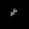 43523MC M: map data used to model this data C: citing same article ( |
|---|---|
| Similar structure data |
-Links
 PDBj
PDBj


- Assembly
| Deposited unit | 
|
|---|---|
| 1 |
|
-Components
-Isoform 4 of Adhesion G protein-coupled receptor ... , 2 types, 2 molecules AB
| #1: Protein | Mass: 41641.762 Da / Num. of mol.: 1 Source method: isolated from a genetically manipulated source Source: (gene. exp.) Homo sapiens (human) / Gene: ADGRL3, KIAA0768, LEC3, LPHN3 / Production host:   Spodoptera frugiperda (fall armyworm) / References: UniProt: Q9HAR2 Spodoptera frugiperda (fall armyworm) / References: UniProt: Q9HAR2 |
|---|---|
| #2: Protein | Mass: 35839.250 Da / Num. of mol.: 1 Source method: isolated from a genetically manipulated source Source: (gene. exp.) Homo sapiens (human) / Gene: ADGRL3, KIAA0768, LEC3, LPHN3 / Production host: Spodoptera frugiperda (fall armyworm) / References: UniProt: Q9HAR2 |
-Antibody , 2 types, 2 molecules CD
| #3: Antibody | Mass: 23329.863 Da / Num. of mol.: 1 Source method: isolated from a genetically manipulated source Source: (gene. exp.) synthetic construct (others) / Production host:  |
|---|---|
| #4: Antibody | Mass: 25221.016 Da / Num. of mol.: 1 Source method: isolated from a genetically manipulated source Source: (gene. exp.) synthetic construct (others) / Production host: |
-Sugars , 2 types, 2 molecules 
| #5: Polysaccharide | beta-D-mannopyranose-(1-4)-2-acetamido-2-deoxy-beta-D-glucopyranose-(1-4)-2-acetamido-2-deoxy-beta- ...beta-D-mannopyranose-(1-4)-2-acetamido-2-deoxy-beta-D-glucopyranose-(1-4)-2-acetamido-2-deoxy-beta-D-glucopyranose Source method: isolated from a genetically manipulated source |
|---|---|
| #6: Sugar | ChemComp-NAG /  Type: D-saccharide, beta linking / Mass: 221.208 Da / Num. of mol.: 1 / Source method: obtained synthetically / Formula: C8H15NO6 Type: D-saccharide, beta linking / Mass: 221.208 Da / Num. of mol.: 1 / Source method: obtained synthetically / Formula: C8H15NO6 |
-Details
| Has ligand of interest | N |
|---|---|
| Has protein modification | Y |
-Experimental details
-Experiment
| Experiment | Method: ELECTRON MICROSCOPY |
|---|---|
| EM experiment | Aggregation state: PARTICLE / 3D reconstruction method: single particle reconstruction |
- Sample preparation
| Component |
| ||||||||||||||||||||||||
|---|---|---|---|---|---|---|---|---|---|---|---|---|---|---|---|---|---|---|---|---|---|---|---|---|---|
| Molecular weight |
| ||||||||||||||||||||||||
| Source (natural) |
| ||||||||||||||||||||||||
| Source (recombinant) |
| ||||||||||||||||||||||||
| Buffer solution | pH: 7.5 | ||||||||||||||||||||||||
| Buffer component |
| ||||||||||||||||||||||||
| Specimen | Conc.: 2.5 mg/ml / Embedding applied: NO / Shadowing applied: NO / Staining applied: NO / Vitrification applied: YES / Details: Monodisperse protein sample in detergent buffer | ||||||||||||||||||||||||
| Specimen support | Grid material: GOLD / Grid mesh size: 300 divisions/in. / Grid type: Quantifoil R1.2/1.3 | ||||||||||||||||||||||||
| Vitrification | Instrument: FEI VITROBOT MARK IV / Cryogen name: ETHANE / Humidity: 100 % / Chamber temperature: 283.15 K |
- Electron microscopy imaging
| Experimental equipment |  Model: Titan Krios / Image courtesy: FEI Company |
|---|---|
| Microscopy | Model: FEI TITAN KRIOS |
| Electron gun | Electron source:  FIELD EMISSION GUN / Accelerating voltage: 300 kV / Illumination mode: OTHER FIELD EMISSION GUN / Accelerating voltage: 300 kV / Illumination mode: OTHER |
| Electron lens | Mode: BRIGHT FIELD / Nominal defocus max: 2000 nm / Nominal defocus min: 1000 nm |
| Image recording | Electron dose: 1.1 e/Å2 / Film or detector model: GATAN K3 (6k x 4k) |
- Processing
| EM software |
| ||||||||||||||||||||||||
|---|---|---|---|---|---|---|---|---|---|---|---|---|---|---|---|---|---|---|---|---|---|---|---|---|---|
| CTF correction | Type: PHASE FLIPPING AND AMPLITUDE CORRECTION | ||||||||||||||||||||||||
| Particle selection | Num. of particles selected: 2621532 | ||||||||||||||||||||||||
| 3D reconstruction | Resolution: 3.9 Å / Resolution method: FSC 0.143 CUT-OFF / Num. of particles: 96958 / Symmetry type: POINT | ||||||||||||||||||||||||
| Refine LS restraints |
|
