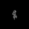
 Movie
Movie Controller


+ Open data

- Basic information
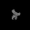
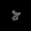
| Entry | Database: PDB / ID: 8vs6 | ||||||
|---|---|---|---|---|---|---|---|
| Title | L-TGF-b3/avb8 | ||||||
 Components Components |
| ||||||
 Keywords Keywords | SIGNALING PROTEIN / TGFb / Complex | ||||||
| Function / homology |  Function and homology information Function and homology informationganglioside metabolic process / uterine wall breakdown / detection of hypoxia / Langerhans cell differentiation / memory T cell differentiation / type III transforming growth factor beta receptor binding / negative regulation of macrophage cytokine production / placenta blood vessel development / integrin alphav-beta8 complex / integrin alphav-beta6 complex ...ganglioside metabolic process / uterine wall breakdown / detection of hypoxia / Langerhans cell differentiation / memory T cell differentiation / type III transforming growth factor beta receptor binding / negative regulation of macrophage cytokine production / placenta blood vessel development / integrin alphav-beta8 complex / integrin alphav-beta6 complex / transforming growth factor beta production / negative regulation of entry of bacterium into host cell / integrin alphav-beta5 complex / secondary palate development / opsonin binding / integrin alphav-beta1 complex / positive regulation of tight junction disassembly / Cross-presentation of particulate exogenous antigens (phagosomes) / extracellular matrix protein binding / type II transforming growth factor beta receptor binding / Laminin interactions / integrin alphav-beta3 complex / negative regulation of lipoprotein metabolic process / cell-cell junction organization / alphav-beta3 integrin-PKCalpha complex / positive regulation of small GTPase mediated signal transduction / entry into host cell by a symbiont-containing vacuole / alphav-beta3 integrin-HMGB1 complex / type I transforming growth factor beta receptor binding / negative regulation of lipid transport / hard palate development / regulation of phagocytosis / mammary gland development / Elastic fibre formation / alphav-beta3 integrin-IGF-1-IGF1R complex / transforming growth factor beta binding / odontogenesis / filopodium membrane / extracellular matrix binding / lung alveolus development / face morphogenesis / cartilage development / apolipoprotein A-I-mediated signaling pathway / negative regulation of low-density lipoprotein particle clearance / wound healing, spreading of epidermal cells / apoptotic cell clearance / positive regulation of filopodium assembly / integrin complex / cell adhesion mediated by integrin / Molecules associated with elastic fibres / heterotypic cell-cell adhesion / negative chemotaxis / positive regulation of osteoblast proliferation / Mechanical load activates signaling by PIEZO1 and integrins in osteocytes / Syndecan interactions / endodermal cell differentiation / microvillus membrane / cell-substrate adhesion / PECAM1 interactions / TGF-beta receptor signaling activates SMADs / positive regulation of collagen biosynthetic process / lamellipodium membrane / fibronectin binding / positive regulation of cell division / positive regulation of SMAD protein signal transduction / positive regulation of intracellular signal transduction / negative regulation of vascular associated smooth muscle cell proliferation / negative regulation of macrophage derived foam cell differentiation / negative regulation of lipid storage / ECM proteoglycans / Integrin cell surface interactions / vasculogenesis / ERK1 and ERK2 cascade / positive regulation of epithelial to mesenchymal transition / salivary gland morphogenesis / specific granule membrane / coreceptor activity / extrinsic apoptotic signaling pathway in absence of ligand / phagocytic vesicle / substrate adhesion-dependent cell spreading / positive regulation of stress fiber assembly / response to progesterone / transforming growth factor beta receptor signaling pathway / positive regulation of cell adhesion / platelet alpha granule lumen / Turbulent (oscillatory, disturbed) flow shear stress activates signaling by PIEZO1 and integrins in endothelial cells / cytokine activity / integrin-mediated signaling pathway / cell-matrix adhesion / negative regulation of extrinsic apoptotic signaling pathway / protein kinase C binding / Signal transduction by L1 / positive regulation of protein secretion / growth factor activity / cell-cell adhesion / VEGFA-VEGFR2 Pathway / integrin binding / ruffle membrane / response to virus / calcium ion transmembrane transport Similarity search - Function | ||||||
| Biological species |  Homo sapiens (human) Homo sapiens (human) | ||||||
| Method | ELECTRON MICROSCOPY / single particle reconstruction / cryo EM / Resolution: 2.73 Å | ||||||
 Authors Authors | Jin, M. / Cheng, Y. / Nishimura, S.L. | ||||||
| Funding support |  United States, 1items United States, 1items
| ||||||
 Citation Citation | Journal: Cell / Year: 2024 Title: Dynamic allostery drives autocrine and paracrine TGF-β signaling. Authors: Mingliang Jin / Robert I Seed / Guoqing Cai / Tiffany Shing / Li Wang / Saburo Ito / Anthony Cormier / Stephanie A Wankowicz / Jillian M Jespersen / Jody L Baron / Nicholas D Carey / Melody ...Authors: Mingliang Jin / Robert I Seed / Guoqing Cai / Tiffany Shing / Li Wang / Saburo Ito / Anthony Cormier / Stephanie A Wankowicz / Jillian M Jespersen / Jody L Baron / Nicholas D Carey / Melody G Campbell / Zanlin Yu / Phu K Tang / Pilar Cossio / Weihua Wen / Jianlong Lou / James Marks / Stephen L Nishimura / Yifan Cheng / Abstract: TGF-β, essential for development and immunity, is expressed as a latent complex (L-TGF-β) non-covalently associated with its prodomain and presented on immune cell surfaces by covalent association ...TGF-β, essential for development and immunity, is expressed as a latent complex (L-TGF-β) non-covalently associated with its prodomain and presented on immune cell surfaces by covalent association with GARP. Binding to integrin αvβ8 activates L-TGF-β1/GARP. The dogma is that mature TGF-β must physically dissociate from L-TGF-β1 for signaling to occur. Our previous studies discovered that αvβ8-mediated TGF-β autocrine signaling can occur without TGF-β1 release from its latent form. Here, we show that mice engineered to express TGF-β1 that cannot release from L-TGF-β1 survive without early lethal tissue inflammation, unlike those with TGF-β1 deficiency. Combining cryogenic electron microscopy with cell-based assays, we reveal a dynamic allosteric mechanism of autocrine TGF-β1 signaling without release where αvβ8 binding redistributes the intrinsic flexibility of L-TGF-β1 to expose TGF-β1 to its receptors. Dynamic allostery explains the TGF-β3 latency/activation mechanism and why TGF-β3 functions distinctly from TGF-β1, suggesting that it broadly applies to other flexible cell surface receptor/ligand systems. | ||||||
| History |
|
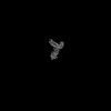
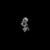
- Structure visualization
| Structure viewer | Molecule: MolmilJmol/JSmol |
|---|
- Downloads & links
-Download
| PDBx/mmCIF format | 8vs6.cif.gz | 337.5 KB | Display | PDBx/mmCIF format |
|---|---|---|---|---|
| PDB format | pdb8vs6.ent.gz | 259.5 KB | Display | PDB format |
| PDBx/mmJSON format | 8vs6.json.gz | Tree view | PDBx/mmJSON format | |
| Others |  Other downloads Other downloads |
-Validation report
| Arichive directory | https://data.pdbj.org/pub/pdb/validation_reports/vs/8vs6ftp://data.pdbj.org/pub/pdb/validation_reports/vs/8vs6 | HTTPS FTP |
|---|
-Related structure data
| Related structure data | 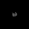 43489MC 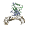 8vsbC  8vscC  8vsdC M: map data used to model this data C: citing same article ( |
|---|---|
| Similar structure data |

-Links
 PDBj
PDBj


- Assembly
| Deposited unit | 
|
|---|---|
| 1 |
|
-Components
-Protein , 3 types, 3 molecules EAB
| #1: Protein | Mass: 44858.094 Da / Num. of mol.: 1 Source method: isolated from a genetically manipulated source Source: (gene. exp.) Homo sapiens (human) / Gene: TGFB3 / Production host: Homo sapiens (human) / References: UniProt: P10600 |
|---|---|
| #2: Protein | Mass: 106375.805 Da / Num. of mol.: 1 Source method: isolated from a genetically manipulated source Source: (gene. exp.) Homo sapiens (human) / Gene: ITGAV, MSK8, VNRA, VTNR / Production host:   Cricetulus griseus (Chinese hamster) / References: UniProt: P06756 Cricetulus griseus (Chinese hamster) / References: UniProt: P06756 |
| #3: Protein | Mass: 71270.758 Da / Num. of mol.: 1 Source method: isolated from a genetically manipulated source Source: (gene. exp.) Homo sapiens (human) / Gene: ITGB8 / Production host: Cricetulus griseus (Chinese hamster) / References: UniProt: P26012 |
-Sugars , 3 types, 8 molecules 
| #4: Polysaccharide | alpha-D-mannopyranose-(1-2)-alpha-D-mannopyranose-(1-3)-[alpha-D-mannopyranose-(1-6)]beta-D- ...alpha-D-mannopyranose-(1-2)-alpha-D-mannopyranose-(1-3)-[alpha-D-mannopyranose-(1-6)]beta-D-mannopyranose-(1-4)-2-acetamido-2-deoxy-beta-D-glucopyranose-(1-4)-2-acetamido-2-deoxy-beta-D-glucopyranose Source method: isolated from a genetically manipulated source | ||
|---|---|---|---|
| #5: Polysaccharide | 2-acetamido-2-deoxy-beta-D-glucopyranose-(1-4)-2-acetamido-2-deoxy-beta-D-glucopyranose Source method: isolated from a genetically manipulated source #6: Sugar |  Type: D-saccharide, beta linking / Mass: 221.208 Da / Num. of mol.: 3 / Source method: obtained synthetically / Formula: C8H15NO6 Type: D-saccharide, beta linking / Mass: 221.208 Da / Num. of mol.: 3 / Source method: obtained synthetically / Formula: C8H15NO6 |
-Non-polymers , 2 types, 6 molecules 

| #7: Chemical | ChemComp-CA /  Mass: 40.078 Da / Num. of mol.: 5 / Source method: obtained synthetically / Formula: Ca Mass: 40.078 Da / Num. of mol.: 5 / Source method: obtained synthetically / Formula: Ca#8: Chemical | ChemComp-MG / |  Mass: 24.305 Da / Num. of mol.: 1 / Source method: obtained synthetically / Formula: Mg Mass: 24.305 Da / Num. of mol.: 1 / Source method: obtained synthetically / Formula: Mg |
|---|
-Details
| Has ligand of interest | N |
|---|---|
| Has protein modification | Y |
-Experimental details
-Experiment
| Experiment | Method: ELECTRON MICROSCOPY |
|---|---|
| EM experiment | Aggregation state: PARTICLE / 3D reconstruction method: single particle reconstruction |
- Sample preparation
| Component |
| ||||||||||||||||||||||||
|---|---|---|---|---|---|---|---|---|---|---|---|---|---|---|---|---|---|---|---|---|---|---|---|---|---|
| Molecular weight |
| ||||||||||||||||||||||||
| Source (natural) |
| ||||||||||||||||||||||||
| Source (recombinant) |
| ||||||||||||||||||||||||
| Buffer solution | pH: 7.4 | ||||||||||||||||||||||||
| Specimen | Embedding applied: NO / Shadowing applied: NO / Staining applied: NO / Vitrification applied: YES | ||||||||||||||||||||||||
| Vitrification | Cryogen name: ETHANE |
- Electron microscopy imaging
| Experimental equipment |  Model: Titan Krios / Image courtesy: FEI Company |
|---|---|
| Microscopy | Model: FEI TITAN KRIOS |
| Electron gun | Electron source:  FIELD EMISSION GUN / Accelerating voltage: 300 kV / Illumination mode: FLOOD BEAM FIELD EMISSION GUN / Accelerating voltage: 300 kV / Illumination mode: FLOOD BEAM |
| Electron lens | Mode: BRIGHT FIELD / Nominal defocus max: 2400 nm / Nominal defocus min: 1200 nm |
| Image recording | Electron dose: 70 e/Å2 / Film or detector model: GATAN K2 SUMMIT (4k x 4k) |
- Processing
| CTF correction | Type: PHASE FLIPPING AND AMPLITUDE CORRECTION | ||||||||||||||||||||||||
|---|---|---|---|---|---|---|---|---|---|---|---|---|---|---|---|---|---|---|---|---|---|---|---|---|---|
| 3D reconstruction | Resolution: 2.73 Å / Resolution method: FSC 0.143 CUT-OFF / Num. of particles: 382107 / Symmetry type: POINT | ||||||||||||||||||||||||
| Refine LS restraints |
|
