[English] 日本語
 Yorodumi
- PDB-8vjo: Cryo-EM structure of Myxococcus xanthus EncA encapsulin shell loa... -
+ Open data
- Basic information
| Entry | Database: PDB / ID: 8vjo | |||||||||||||||||||||||||||||||||||||||||||||||||||||||||||||||||||||
|---|---|---|---|---|---|---|---|---|---|---|---|---|---|---|---|---|---|---|---|---|---|---|---|---|---|---|---|---|---|---|---|---|---|---|---|---|---|---|---|---|---|---|---|---|---|---|---|---|---|---|---|---|---|---|---|---|---|---|---|---|---|---|---|---|---|---|---|---|---|---|
| Title | Cryo-EM structure of Myxococcus xanthus EncA encapsulin shell loaded with EncD cargo | |||||||||||||||||||||||||||||||||||||||||||||||||||||||||||||||||||||
 Components Components |
| |||||||||||||||||||||||||||||||||||||||||||||||||||||||||||||||||||||
 Keywords Keywords | VIRUS LIKE PARTICLE / Encapsulin / EncA / EncD / flavin / flavin mononucleotide / iron / ferroxidase / ferritin / ferric reductase | |||||||||||||||||||||||||||||||||||||||||||||||||||||||||||||||||||||
| Function / homology | Type 1 encapsulin shell protein / Encapsulating protein for peroxidase / : / encapsulin nanocompartment / iron ion transport / intracellular iron ion homeostasis / metal ion binding / Type 1 encapsulin shell protein EncA / Encapsulin nanocompartment cargo protein EncD Function and homology information Function and homology information | |||||||||||||||||||||||||||||||||||||||||||||||||||||||||||||||||||||
| Biological species |  Myxococcus xanthus (bacteria) Myxococcus xanthus (bacteria) | |||||||||||||||||||||||||||||||||||||||||||||||||||||||||||||||||||||
| Method | ELECTRON MICROSCOPY / single particle reconstruction / cryo EM / Resolution: 2.4 Å | |||||||||||||||||||||||||||||||||||||||||||||||||||||||||||||||||||||
 Authors Authors | Eren, E. | |||||||||||||||||||||||||||||||||||||||||||||||||||||||||||||||||||||
| Funding support |  United States, 1items United States, 1items
| |||||||||||||||||||||||||||||||||||||||||||||||||||||||||||||||||||||
 Citation Citation |  Journal: Proc Natl Acad Sci U S A / Year: 2024 Journal: Proc Natl Acad Sci U S A / Year: 2024Title: encapsulin cargo protein EncD is a flavin-binding protein with ferric reductase activity. Authors: Elif Eren / Norman R Watts / James F Conway / Paul T Wingfield /  Abstract: Encapsulins are protein nanocompartments that regulate cellular metabolism in several bacteria and archaea. encapsulins protect the bacterial cells against oxidative stress by sequestering cytosolic ...Encapsulins are protein nanocompartments that regulate cellular metabolism in several bacteria and archaea. encapsulins protect the bacterial cells against oxidative stress by sequestering cytosolic iron. These encapsulins are formed by the shell protein EncA and three cargo proteins: EncB, EncC, and EncD. EncB and EncC form rotationally symmetric decamers with ferroxidase centers (FOCs) that oxidize Fe to Fe for iron storage in mineral form. However, the structure and function of the third cargo protein, EncD, have yet to be determined. Here, we report the x-ray crystal structure of EncD in complex with flavin mononucleotide. EncD forms an α-helical hairpin arranged as an antiparallel dimer, but unlike other flavin-binding proteins, it has no β-sheet, showing that EncD and its homologs represent a unique class of bacterial flavin-binding proteins. The cryo-EM structure of EncA-EncD encapsulins confirms that EncD binds to the interior of the EncA shell via its C-terminal targeting peptide. With only 100 amino acids, the EncD α-helical dimer forms the smallest flavin-binding domain observed to date. Unlike EncB and EncC, EncD lacks a FOC, and our biochemical results show that EncD instead is a NAD(P)H-dependent ferric reductase, indicating that the encapsulins act as an integrated system for iron homeostasis. Overall, this work contributes to our understanding of bacterial metabolism and could lead to the development of technologies for iron biomineralization and the production of iron-containing materials for the treatment of various diseases associated with oxidative stress. | |||||||||||||||||||||||||||||||||||||||||||||||||||||||||||||||||||||
| History |
|
- Structure visualization
| Structure viewer | Molecule:  Molmil Molmil Jmol/JSmol Jmol/JSmol |
|---|
- Downloads & links
- Download
| PDBx/mmCIF format |  8vjo.cif.gz 8vjo.cif.gz | 157.1 KB | Display |  PDBx/mmCIF format PDBx/mmCIF format |
|---|---|---|---|---|
| PDB format |  pdb8vjo.ent.gz pdb8vjo.ent.gz | 124 KB | Display |  PDB format PDB format |
| PDBx/mmJSON format |  8vjo.json.gz 8vjo.json.gz | Tree view |  PDBx/mmJSON format PDBx/mmJSON format | |
| Others |  Other downloads Other downloads |
-Validation report
| Summary document |  8vjo_validation.pdf.gz 8vjo_validation.pdf.gz | 1.4 MB | Display |  wwPDB validaton report wwPDB validaton report |
|---|---|---|---|---|
| Full document |  8vjo_full_validation.pdf.gz 8vjo_full_validation.pdf.gz | 1.4 MB | Display | |
| Data in XML |  8vjo_validation.xml.gz 8vjo_validation.xml.gz | 48.1 KB | Display | |
| Data in CIF |  8vjo_validation.cif.gz 8vjo_validation.cif.gz | 71.4 KB | Display | |
| Arichive directory |  https://data.pdbj.org/pub/pdb/validation_reports/vj/8vjo https://data.pdbj.org/pub/pdb/validation_reports/vj/8vjo ftp://data.pdbj.org/pub/pdb/validation_reports/vj/8vjo ftp://data.pdbj.org/pub/pdb/validation_reports/vj/8vjo | HTTPS FTP |
-Related structure data
| Related structure data |  43290MC  8vjnC C: citing same article ( M: map data used to model this data |
|---|---|
| Similar structure data | Similarity search - Function & homology  F&H Search F&H Search |
- Links
Links
- Assembly
| Deposited unit | 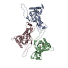
|
|---|---|
| 1 | x 60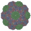
|
| 2 |
|
| 3 | x 5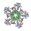
|
| 4 | x 6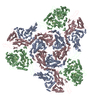
|
| 5 | 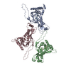
|
| Symmetry | Point symmetry: (Schoenflies symbol: I (icosahedral)) |
- Components
| #1: Protein | Mass: 33505.074 Da / Num. of mol.: 3 Source method: isolated from a genetically manipulated source Source: (gene. exp.)  Myxococcus xanthus (bacteria) / Gene: MXAN_3556 / Production host: Myxococcus xanthus (bacteria) / Gene: MXAN_3556 / Production host:  #2: Protein/peptide | Mass: 859.992 Da / Num. of mol.: 3 Fragment: targeting peptide: residues 106-114 (99-107 in Uniprot numbering) Source method: isolated from a genetically manipulated source Source: (gene. exp.)  Myxococcus xanthus (bacteria) / Gene: encD, MXAN_2410 / Production host: Myxococcus xanthus (bacteria) / Gene: encD, MXAN_2410 / Production host:  Has protein modification | N | |
|---|
-Experimental details
-Experiment
| Experiment | Method: ELECTRON MICROSCOPY |
|---|---|
| EM experiment | Aggregation state: PARTICLE / 3D reconstruction method: single particle reconstruction |
- Sample preparation
| Component | Name: Icosahedral encapsulin EncA particles in complex with cargo protein EncD Type: ORGANELLE OR CELLULAR COMPONENT / Entity ID: all / Source: RECOMBINANT |
|---|---|
| Molecular weight | Experimental value: NO |
| Source (natural) | Organism:  Myxococcus xanthus (bacteria) Myxococcus xanthus (bacteria) |
| Source (recombinant) | Organism:  |
| Buffer solution | pH: 7.3 / Details: 20mM HEPES, 150mM NaCl |
| Specimen | Conc.: 0.5 mg/ml / Embedding applied: NO / Shadowing applied: NO / Staining applied: NO / Vitrification applied: YES |
| Specimen support | Grid material: COPPER / Grid mesh size: 300 divisions/in. / Grid type: Quantifoil R2/1 |
| Vitrification | Instrument: FEI VITROBOT MARK IV / Cryogen name: ETHANE |
- Electron microscopy imaging
| Experimental equipment |  Model: Titan Krios / Image courtesy: FEI Company |
|---|---|
| Microscopy | Model: FEI TITAN KRIOS |
| Electron gun | Electron source:  FIELD EMISSION GUN / Accelerating voltage: 300 kV / Illumination mode: FLOOD BEAM FIELD EMISSION GUN / Accelerating voltage: 300 kV / Illumination mode: FLOOD BEAM |
| Electron lens | Mode: BRIGHT FIELD / Nominal magnification: 130000 X / Nominal defocus max: 2600 nm / Nominal defocus min: 600 nm |
| Image recording | Electron dose: 50 e/Å2 / Film or detector model: FEI FALCON IV (4k x 4k) |
- Processing
| EM software |
| ||||||||||||||||||||||||||||
|---|---|---|---|---|---|---|---|---|---|---|---|---|---|---|---|---|---|---|---|---|---|---|---|---|---|---|---|---|---|
| CTF correction | Type: PHASE FLIPPING AND AMPLITUDE CORRECTION | ||||||||||||||||||||||||||||
| Symmetry | Point symmetry: I (icosahedral) | ||||||||||||||||||||||||||||
| 3D reconstruction | Resolution: 2.4 Å / Resolution method: FSC 0.143 CUT-OFF / Num. of particles: 47437 / Num. of class averages: 1 / Symmetry type: POINT | ||||||||||||||||||||||||||||
| Refine LS restraints |
|
 Movie
Movie Controller


 PDBj
PDBj

