 Movie
Movie Controller


+ Open data

- Basic information
| Entry | Database: PDB / ID: 8v02 | |||||||||
|---|---|---|---|---|---|---|---|---|---|---|
| Title | AaegOR10 structure bound to o-cresol | |||||||||
 Components Components |
| |||||||||
 Keywords Keywords | MEMBRANE PROTEIN / Mosquito olfactory receptor OR10 | |||||||||
| Function / homology |  Function and homology information Function and homology informationolfactory receptor activity / odorant binding / signal transduction / identical protein binding / plasma membrane Similarity search - Function | |||||||||
| Biological species |  Apocrypta bakeri (insect) Apocrypta bakeri (insect) | |||||||||
| Method | ELECTRON MICROSCOPY / single particle reconstruction / cryo EM / Resolution: 2.9 Å | |||||||||
 Authors Authors | Zhao, J. / del Marmol, J. | |||||||||
| Funding support |  United States, 2items United States, 2items
| |||||||||
 Citation Citation | Journal: Science / Year: 2024 Title: Structural basis of odor sensing by insect heteromeric odorant receptors. Authors: Jiawei Zhao / Andy Q Chen / Jaewook Ryu / Josefina Del Mármol / Abstract: Most insects, including human-targeting mosquitoes, detect odors through odorant-activated ion channel complexes consisting of a divergent odorant-binding subunit (OR) and a conserved co-receptor ...Most insects, including human-targeting mosquitoes, detect odors through odorant-activated ion channel complexes consisting of a divergent odorant-binding subunit (OR) and a conserved co-receptor subunit (Orco). As a basis for understanding how odorants activate these heteromeric receptors, we report here cryo-electron microscopy structures of two different heteromeric odorant receptor complexes containing ORs from disease-vector mosquitos or . These structures reveal an unexpected stoichiometry of one OR to three Orco subunits. Comparison of structures in odorant-bound and unbound states indicates that odorant binding to the sole OR subunit is sufficient to open the channel pore, suggesting a mechanism of OR activation and a conceptual framework for understanding evolution of insect odorant receptor sensitivity. | |||||||||
| History |
|
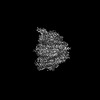
- Structure visualization
| Structure viewer | Molecule: MolmilJmol/JSmol |
|---|
- Downloads & links
-Download
| PDBx/mmCIF format | 8v02.cif.gz | 722 KB | Display | PDBx/mmCIF format |
|---|---|---|---|---|
| PDB format | pdb8v02.ent.gz | 484.7 KB | Display | PDB format |
| PDBx/mmJSON format | 8v02.json.gz | Tree view | PDBx/mmJSON format | |
| Others |  Other downloads Other downloads |
-Validation report
| Arichive directory | https://data.pdbj.org/pub/pdb/validation_reports/v0/8v02ftp://data.pdbj.org/pub/pdb/validation_reports/v0/8v02 | HTTPS FTP |
|---|
-Related structure data
| Related structure data |  42850MC 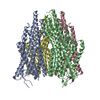 8v00C 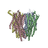 8v3cC 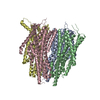 8v3dC M: map data used to model this data C: citing same article ( |
|---|---|
| Similar structure data |

-Links
 PDBj
PDBj- Assembly
| Deposited unit | 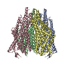
|
|---|---|
| 1 |
|
-Components
| #1: Protein | Mass: 43491.168 Da / Num. of mol.: 1 Source method: isolated from a genetically manipulated source Source: (gene. exp.)  Homo sapiens (human) / References: UniProt: Q177X3 Homo sapiens (human) / References: UniProt: Q177X3 | ||||
|---|---|---|---|---|---|
| #2: Protein | Mass: 53204.953 Da / Num. of mol.: 3 Source method: isolated from a genetically manipulated source Source: (gene. exp.) Apocrypta bakeri (insect) / Gene: Or2 / Production host: Homo sapiens (human) / References: UniProt: B0FAQ4#3: Chemical | ChemComp-JZ0 / | 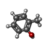  Mass: 108.138 Da / Num. of mol.: 1 / Source method: obtained synthetically / Formula: C7H8O / Feature type: SUBJECT OF INVESTIGATION Mass: 108.138 Da / Num. of mol.: 1 / Source method: obtained synthetically / Formula: C7H8O / Feature type: SUBJECT OF INVESTIGATIONHas ligand of interest | Y | |
-Experimental details
-Experiment
| Experiment | Method: ELECTRON MICROSCOPY |
|---|---|
| EM experiment | Aggregation state: PARTICLE / 3D reconstruction method: single particle reconstruction |
- Sample preparation
| Component |
| ||||||||||||||||||||||||
|---|---|---|---|---|---|---|---|---|---|---|---|---|---|---|---|---|---|---|---|---|---|---|---|---|---|
| Molecular weight |
| ||||||||||||||||||||||||
| Source (natural) |
| ||||||||||||||||||||||||
| Source (recombinant) |
| ||||||||||||||||||||||||
| Buffer solution | pH: 7.5 | ||||||||||||||||||||||||
| Specimen | Embedding applied: NO / Shadowing applied: NO / Staining applied: NO / Vitrification applied: YES | ||||||||||||||||||||||||
| Vitrification | Cryogen name: ETHANE |
- Electron microscopy imaging
| Experimental equipment |  Model: Titan Krios / Image courtesy: FEI Company | ||||||||||||
|---|---|---|---|---|---|---|---|---|---|---|---|---|---|
| Microscopy | Model: FEI TITAN KRIOS | ||||||||||||
| Electron gun | Electron source:  FIELD EMISSION GUN / Accelerating voltage: 300 kV / Illumination mode: FLOOD BEAM FIELD EMISSION GUN / Accelerating voltage: 300 kV / Illumination mode: FLOOD BEAM | ||||||||||||
| Electron lens | Mode: BRIGHT FIELD / Nominal defocus max: 2000 nm / Nominal defocus min: 1000 nm | ||||||||||||
| Image recording |
|
- Processing
| EM software | Name: PHENIX / Version: 1.21_5207 / Category: model refinement | ||||||||||||||||||||||||
|---|---|---|---|---|---|---|---|---|---|---|---|---|---|---|---|---|---|---|---|---|---|---|---|---|---|
| CTF correction | Type: PHASE FLIPPING AND AMPLITUDE CORRECTION | ||||||||||||||||||||||||
| 3D reconstruction | Resolution: 2.9 Å / Resolution method: FSC 0.143 CUT-OFF / Num. of particles: 496871 / Symmetry type: POINT | ||||||||||||||||||||||||
| Refinement | Cross valid method: NONE Stereochemistry target values: GeoStd + Monomer Library + CDL v1.2 | ||||||||||||||||||||||||
| Displacement parameters | Biso mean: 37.89 Å2 | ||||||||||||||||||||||||
| Refine LS restraints |
|
