 Movie
Movie Controller


+ Open data

- Basic information
| Entry | Database: PDB / ID: 8ug5 | ||||||||||||
|---|---|---|---|---|---|---|---|---|---|---|---|---|---|
| Title | Caenorhabditis elegans Otopetrin 8 (CeOtop8) in pH 5.0 | ||||||||||||
 Components Components | Otopetrin-2 | ||||||||||||
 Keywords Keywords | MEMBRANE PROTEIN / Proton channel / Otop / C.elegans / Otopetrin | ||||||||||||
| Function / homology | Otopetrin / Otopetrin / proton channel activity / proton transmembrane transport / plasma membrane / Otopetrin-2 Function and homology information Function and homology information | ||||||||||||
| Biological species |  | ||||||||||||
| Method | ELECTRON MICROSCOPY / single particle reconstruction / cryo EM / Resolution: 2.91 Å | ||||||||||||
 Authors Authors | Gan, N. / Jiang, Y. | ||||||||||||
| Funding support |  United States, 3items United States, 3items
| ||||||||||||
 Citation Citation | Journal: Nat Commun / Year: 2024 Title: Structural mechanism of proton conduction in otopetrin proton channel. Authors: Ninghai Gan / Weizhong Zeng / Yan Han / Qingfeng Chen / Youxing Jiang /  Abstract: The otopetrin (OTOP) proteins were recently characterized as extracellular proton-activated proton channels. Several recent OTOP channel structures demonstrated that the channels form a dimer with ...The otopetrin (OTOP) proteins were recently characterized as extracellular proton-activated proton channels. Several recent OTOP channel structures demonstrated that the channels form a dimer with each subunit adopting a double-barrel architecture. However, the structural mechanisms underlying some basic functional properties of the OTOP channels remain unresolved, including extracellular pH activation, proton conducting pathway, and rapid desensitization. In this study, we performed structural and functional characterization of the Caenorhabditis elegans OTOP8 (CeOTOP8) and mouse OTOP2 (mOTOP2) and illuminated a set of conformational changes related to the proton-conducting process in OTOP. The structures of CeOTOP8 reveal the conformational change at the N-terminal part of TM12 that renders the channel in a transiently proton-transferring state, elucidating an inter-barrel, Glu/His-bridged proton passage within each subunit. The structures of mOTOP2 reveal the conformational change at the N-terminal part of TM6 that exposes the central glutamate to the extracellular solution for protonation. In addition, the structural comparison between CeOTOP8 and mOTOP2, along with the structure-based mutagenesis, demonstrates that an inter-subunit movement at the OTOP channel dimer interface plays a central role in regulating channel activity. Combining the structural information from both channels, we propose a working model describing the multi-step conformational changes during the proton conducting process. | ||||||||||||
| History |
|
- Structure visualization
| Structure viewer | Molecule: MolmilJmol/JSmol |
|---|
- Downloads & links
-Download
| PDBx/mmCIF format | 8ug5.cif.gz | 149.1 KB | Display | PDBx/mmCIF format |
|---|---|---|---|---|
| PDB format | pdb8ug5.ent.gz | 114.2 KB | Display | PDB format |
| PDBx/mmJSON format | 8ug5.json.gz | Tree view | PDBx/mmJSON format | |
| Others |  Other downloads Other downloads |
-Validation report
| Arichive directory | https://data.pdbj.org/pub/pdb/validation_reports/ug/8ug5ftp://data.pdbj.org/pub/pdb/validation_reports/ug/8ug5 | HTTPS FTP |
|---|
-Related structure data
| Related structure data | 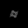 42214MC  8ug4C  8ug6C  8ug7C  8ug8C  8ugaC M: map data used to model this data C: citing same article ( |
|---|---|
| Similar structure data |


-Links
 PDBj
PDBj- Assembly
| Deposited unit | 
|
|---|---|
| 1 |
|
-Components
| #1: Protein | Mass: 69791.508 Da / Num. of mol.: 2 Source method: isolated from a genetically manipulated source Source: (gene. exp.)  Homo sapiens (human) / References: UniProt: G5EDX5 Homo sapiens (human) / References: UniProt: G5EDX5Has protein modification | Y | |
|---|
-Experimental details
-Experiment
| Experiment | Method: ELECTRON MICROSCOPY |
|---|---|
| EM experiment | Aggregation state: PARTICLE / 3D reconstruction method: single particle reconstruction |
- Sample preparation
| Component | Name: CeOTOP8 / Type: COMPLEX / Entity ID: all / Source: RECOMBINANT |
|---|---|
| Source (natural) | Organism: |
| Source (recombinant) | Organism: Homo sapiens (human) |
| Buffer solution | pH: 5 |
| Specimen | Embedding applied: NO / Shadowing applied: NO / Staining applied: NO / Vitrification applied: YES |
| Vitrification | Cryogen name: ETHANE |
- Electron microscopy imaging
| Experimental equipment |  Model: Titan Krios / Image courtesy: FEI Company |
|---|---|
| Microscopy | Model: FEI TITAN KRIOS |
| Electron gun | Electron source:  FIELD EMISSION GUN / Accelerating voltage: 300 kV / Illumination mode: OTHER FIELD EMISSION GUN / Accelerating voltage: 300 kV / Illumination mode: OTHER |
| Electron lens | Mode: DIFFRACTION / Nominal defocus max: 2200 nm / Nominal defocus min: 900 nm |
| Image recording | Electron dose: 60 e/Å2 / Film or detector model: GATAN K3 (6k x 4k) |
- Processing
| CTF correction | Type: PHASE FLIPPING ONLY |
|---|---|
| 3D reconstruction | Resolution: 2.91 Å / Resolution method: FSC 0.143 CUT-OFF / Num. of particles: 41311 / Symmetry type: POINT |
