 Movie
Movie Controller

[English] 日本語
 Yorodumi
- PDB-8uf7: Cryo-EM structure of POmAb, a Type-I anti-prothrombin antiphospho... -
+ Open data

- Basic information
| Entry | Database: PDB / ID: 8uf7 | |||||||||||||||||||||||||||||||||||||||||||||||||||||||||||||||||||||||||||||||||||||||||||||
|---|---|---|---|---|---|---|---|---|---|---|---|---|---|---|---|---|---|---|---|---|---|---|---|---|---|---|---|---|---|---|---|---|---|---|---|---|---|---|---|---|---|---|---|---|---|---|---|---|---|---|---|---|---|---|---|---|---|---|---|---|---|---|---|---|---|---|---|---|---|---|---|---|---|---|---|---|---|---|---|---|---|---|---|---|---|---|---|---|---|---|---|---|---|---|
| Title | Cryo-EM structure of POmAb, a Type-I anti-prothrombin antiphospholipid antibody, bound to kringle-1 of human prothrombin | |||||||||||||||||||||||||||||||||||||||||||||||||||||||||||||||||||||||||||||||||||||||||||||
 Components Components |
| |||||||||||||||||||||||||||||||||||||||||||||||||||||||||||||||||||||||||||||||||||||||||||||
 Keywords Keywords | BLOOD CLOTTING / Autoimmunity / Thrombosis / Complex / Immunoglobulin / Coagulation factor / Anticoagulation / Inhibitor | |||||||||||||||||||||||||||||||||||||||||||||||||||||||||||||||||||||||||||||||||||||||||||||
| Function / homology |  Function and homology information Function and homology information: / thrombospondin receptor activity / thrombin / thrombin-activated receptor signaling pathway / Defective factor XII causes hereditary angioedema / negative regulation of astrocyte differentiation / regulation of blood coagulation / neutrophil-mediated killing of gram-negative bacterium / positive regulation of phospholipase C-activating G protein-coupled receptor signaling pathway / Defective F8 cleavage by thrombin ...: / thrombospondin receptor activity / thrombin / thrombin-activated receptor signaling pathway / Defective factor XII causes hereditary angioedema / negative regulation of astrocyte differentiation / regulation of blood coagulation / neutrophil-mediated killing of gram-negative bacterium / positive regulation of phospholipase C-activating G protein-coupled receptor signaling pathway / Defective F8 cleavage by thrombin / ligand-gated ion channel signaling pathway / Platelet Aggregation (Plug Formation) / positive regulation of collagen biosynthetic process / negative regulation of platelet activation / negative regulation of blood coagulation / negative regulation of fibrinolysis / blood coagulation, fibrin clot formation / positive regulation of blood coagulation / Transport of gamma-carboxylated protein precursors from the endoplasmic reticulum to the Golgi apparatus / : / Gamma-carboxylation of protein precursors / Removal of aminoterminal propeptides from gamma-carboxylated proteins / regulation of cytosolic calcium ion concentration / fibrinolysis / : / negative regulation of proteolysis / negative regulation of cytokine production involved in inflammatory response / Regulation of Complement cascade / positive regulation of release of sequestered calcium ion into cytosol / acute-phase response / Cell surface interactions at the vascular wall / Peptide ligand-binding receptors / growth factor activity / positive regulation of receptor signaling pathway via JAK-STAT / lipopolysaccharide binding / platelet activation / positive regulation of protein localization to nucleus / response to wounding / Golgi lumen / Regulation of Insulin-like Growth Factor (IGF) transport and uptake by Insulin-like Growth Factor Binding Proteins (IGFBPs) / positive regulation of reactive oxygen species metabolic process / blood coagulation / positive regulation of insulin secretion / regulation of cell shape / antimicrobial humoral immune response mediated by antimicrobial peptide / heparin binding / Thrombin signalling through proteinase activated receptors (PARs) / positive regulation of cell growth / blood microparticle / G alpha (q) signalling events / positive regulation of phosphatidylinositol 3-kinase/protein kinase B signal transduction / cell surface receptor signaling pathway / endoplasmic reticulum lumen / receptor ligand activity / serine-type endopeptidase activity / signaling receptor binding / calcium ion binding / positive regulation of cell population proliferation / proteolysis / : / extracellular exosome / extracellular region / plasma membrane Similarity search - Function | |||||||||||||||||||||||||||||||||||||||||||||||||||||||||||||||||||||||||||||||||||||||||||||
| Biological species |   Homo sapiens (human) Homo sapiens (human) | |||||||||||||||||||||||||||||||||||||||||||||||||||||||||||||||||||||||||||||||||||||||||||||
| Method | ELECTRON MICROSCOPY / single particle reconstruction / cryo EM / Resolution: 3.2 Å | |||||||||||||||||||||||||||||||||||||||||||||||||||||||||||||||||||||||||||||||||||||||||||||
 Authors Authors | Kumar, S. / Summers, B. / Basore, K. / Pozzi, N. | |||||||||||||||||||||||||||||||||||||||||||||||||||||||||||||||||||||||||||||||||||||||||||||
| Funding support |  United States, 2items United States, 2items
| |||||||||||||||||||||||||||||||||||||||||||||||||||||||||||||||||||||||||||||||||||||||||||||
 Citation Citation | Journal: Blood / Year: 2024 Title: Cryo-EM structure and functional basis of prothrombin recognition by a type I antiprothrombin antiphospholipid antibody. Authors: Suresh Kumar / Brock Summers / Kathrine Basore / Vittorio Pengo / Robert Flaumenhaft / Nicola Pozzi /  Abstract: Antiprothrombin antibodies are found in antiphospholipid patients, but how they interact with prothrombin remains elusive. Prothrombin adopts closed and open forms. We recently discovered type I and ...Antiprothrombin antibodies are found in antiphospholipid patients, but how they interact with prothrombin remains elusive. Prothrombin adopts closed and open forms. We recently discovered type I and type II antibodies and proposed that type I recognizes the open form. In this study, we report the discovery and structural and functional characterization in human plasma of a type I antibody, POmAb (prothrombin open monoclonal antibody). Using surface plasmon resonance and single-molecule spectroscopy, we show that POmAb interacts with kringle-1 of prothrombin, shifting the equilibrium toward the open form. Using single-particle cryogenic electron microscopy (cryo-EM), we establish that the epitope targeted by POmAb is in kringle-1, comprising an extended binding interface centered at residues R90-Y93. The 3.2-Å cryo-EM structure of the complex reveals that the epitope overlaps with the position occupied by the protease domain of prothrombin in the closed state, explaining the exclusive binding of POmAb to the open form. In human plasma, POmAb prolongs phospholipid-initiated and diluted Russell's viper venom clotting time, which could be partly rescued by excess phospholipids, indicating POmAb is an anticoagulant but exerts a weak lupus anticoagulant effect. These studies reveal the structural basis of prothrombin recognition by a type I antiphospholipid antibody and uncover an exciting new strategy to achieve anticoagulation in human plasma. | |||||||||||||||||||||||||||||||||||||||||||||||||||||||||||||||||||||||||||||||||||||||||||||
| History |
|
- Structure visualization
| Structure viewer | Molecule: MolmilJmol/JSmol |
|---|
- Downloads & links
-Download
| PDBx/mmCIF format | 8uf7.cif.gz | 121.7 KB | Display | PDBx/mmCIF format |
|---|---|---|---|---|
| PDB format | pdb8uf7.ent.gz | 85.4 KB | Display | PDB format |
| PDBx/mmJSON format | 8uf7.json.gz | Tree view | PDBx/mmJSON format | |
| Others |  Other downloads Other downloads |
-Validation report
| Arichive directory | https://data.pdbj.org/pub/pdb/validation_reports/uf/8uf7ftp://data.pdbj.org/pub/pdb/validation_reports/uf/8uf7 | HTTPS FTP |
|---|
-Related structure data
| Related structure data |  42185MC M: map data used to model this data C: citing same article ( |
|---|---|
| Similar structure data |
-Links
 PDBj
PDBj


- Assembly
| Deposited unit | 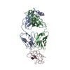
|
|---|---|
| 1 |
|
-Components
| #1: Antibody | Mass: 23558.154 Da / Num. of mol.: 1 Source method: isolated from a genetically manipulated source Source: (gene. exp.)  Cricetulus griseus (Chinese hamster) Cricetulus griseus (Chinese hamster) |
|---|---|
| #2: Antibody | Mass: 23406.252 Da / Num. of mol.: 1 Source method: isolated from a genetically manipulated source Source: (gene. exp.) Cricetulus griseus (Chinese hamster) |
| #3: Protein | Mass: 65386.113 Da / Num. of mol.: 1 / Source method: isolated from a natural source / Source: (natural) Homo sapiens (human) / References: UniProt: P00734 |
| Has ligand of interest | N |
| Has protein modification | Y |
-Experimental details
-Experiment
| Experiment | Method: ELECTRON MICROSCOPY |
|---|---|
| EM experiment | Aggregation state: PARTICLE / 3D reconstruction method: single particle reconstruction |
- Sample preparation
| Component | Name: Complex of Fab fragment of POmAb with human prothrombin Type: COMPLEX Details: Fab fragment generated by proteolytic cleavage of POmAb IgG antibody in complex with open conformation of human prothrombin Entity ID: all / Source: MULTIPLE SOURCES |
|---|---|
| Molecular weight | Value: 0.12 MDa / Experimental value: YES |
| Source (natural) | Organism: |
| Source (recombinant) | Organism: Cricetulus griseus (Chinese hamster) |
| Buffer solution | pH: 7.4 |
| Specimen | Conc.: 0.33 mg/ml / Embedding applied: NO / Shadowing applied: NO / Staining applied: NO / Vitrification applied: YES / Details: Monodisperse particles. Complex purified by SEC. |
| Specimen support | Grid type: Quantifoil R2/2 |
| Vitrification | Instrument: FEI VITROBOT MARK IV / Cryogen name: ETHANE / Humidity: 95 % / Chamber temperature: 277 K |
- Electron microscopy imaging
| Experimental equipment |  Model: Titan Krios / Image courtesy: FEI Company |
|---|---|
| Microscopy | Model: FEI TITAN KRIOS |
| Electron gun | Electron source:  FIELD EMISSION GUN / Accelerating voltage: 300 kV / Illumination mode: FLOOD BEAM FIELD EMISSION GUN / Accelerating voltage: 300 kV / Illumination mode: FLOOD BEAM |
| Electron lens | Mode: BRIGHT FIELD / Nominal magnification: 59000 X / Nominal defocus max: 2400 nm / Nominal defocus min: 1000 nm / Cs: 0.01 mm / Alignment procedure: ZEMLIN TABLEAU |
| Specimen holder | Cryogen: NITROGEN / Specimen holder model: FEI TITAN KRIOS AUTOGRID HOLDER / Temperature (max): 84 K / Temperature (min): 82 K |
| Image recording | Electron dose: 62.2 e/Å2 / Film or detector model: FEI FALCON IV (4k x 4k) |
| EM imaging optics | Spherical aberration corrector: Microscope is outfitted with a Cs image corrector with two hexapole elements. |
| Image scans | Width: 4096 / Height: 4096 |
- Processing
| EM software |
| ||||||||||||||||||||||||
|---|---|---|---|---|---|---|---|---|---|---|---|---|---|---|---|---|---|---|---|---|---|---|---|---|---|
| CTF correction | Type: PHASE FLIPPING AND AMPLITUDE CORRECTION | ||||||||||||||||||||||||
| Symmetry | Point symmetry: C1 (asymmetric) | ||||||||||||||||||||||||
| 3D reconstruction | Resolution: 3.2 Å / Resolution method: FSC 0.143 CUT-OFF / Num. of particles: 176744 / Symmetry type: POINT | ||||||||||||||||||||||||
| Atomic model building | Space: REAL | ||||||||||||||||||||||||
| Refine LS restraints |
|
