 ムービー
ムービー コントローラー


+ データを開く

- 基本情報
基本情報
| 登録情報 | データベース: PDB / ID: 8uf7 | |||||||||
|---|---|---|---|---|---|---|---|---|---|---|
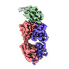
| タイトル | Cryo-EM structure of POmAb, a Type-I anti-prothrombin antiphospholipid antibody, bound to kringle-1 of human prothrombin | |||||||||
 要素 要素 |
| |||||||||
 キーワード キーワード |  BLOOD CLOTTING (凝固・線溶系) / Autoimmunity (自己免疫) / Thrombosis (血栓症) / Complex / Immunoglobulin (抗体) / Coagulation factor (凝固・線溶系) / Anticoagulation / Inhibitor BLOOD CLOTTING (凝固・線溶系) / Autoimmunity (自己免疫) / Thrombosis (血栓症) / Complex / Immunoglobulin (抗体) / Coagulation factor (凝固・線溶系) / Anticoagulation / Inhibitor | |||||||||
| 機能・相同性 |  機能・相同性情報 機能・相同性情報positive regulation of lipid kinase activity / positive regulation of phospholipase C-activating G protein-coupled receptor signaling pathway / cytolysis by host of symbiont cells / thrombospondin receptor activity / Defective factor XII causes hereditary angioedema / トロンビン / neutrophil-mediated killing of gram-negative bacterium / regulation of blood coagulation / ligand-gated ion channel signaling pathway / Defective F8 cleavage by thrombin ...positive regulation of lipid kinase activity / positive regulation of phospholipase C-activating G protein-coupled receptor signaling pathway / cytolysis by host of symbiont cells / thrombospondin receptor activity / Defective factor XII causes hereditary angioedema / トロンビン / neutrophil-mediated killing of gram-negative bacterium / regulation of blood coagulation / ligand-gated ion channel signaling pathway / Defective F8 cleavage by thrombin / Platelet Aggregation (Plug Formation) / negative regulation of astrocyte differentiation / negative regulation of platelet activation / positive regulation of collagen biosynthetic process / negative regulation of cytokine production involved in inflammatory response / positive regulation of blood coagulation / negative regulation of fibrinolysis / Gamma-carboxylation of protein precursors / Transport of gamma-carboxylated protein precursors from the endoplasmic reticulum to the Golgi apparatus / Common Pathway of Fibrin Clot Formation / Removal of aminoterminal propeptides from gamma-carboxylated proteins / regulation of cytosolic calcium ion concentration / fibrinolysis / Intrinsic Pathway of Fibrin Clot Formation / Peptide ligand-binding receptors / positive regulation of release of sequestered calcium ion into cytosol / Regulation of Complement cascade / acute-phase response / Cell surface interactions at the vascular wall / lipopolysaccharide binding / negative regulation of proteolysis / positive regulation of receptor signaling pathway via JAK-STAT / growth factor activity / positive regulation of insulin secretion / 凝固・線溶系 / response to wounding / Golgi lumen / positive regulation of protein localization to nucleus / Regulation of Insulin-like Growth Factor (IGF) transport and uptake by Insulin-like Growth Factor Binding Proteins (IGFBPs) / positive regulation of reactive oxygen species metabolic process / 凝固・線溶系 / antimicrobial humoral immune response mediated by antimicrobial peptide / Thrombin signalling through proteinase activated receptors (PARs) / heparin binding / regulation of cell shape / positive regulation of cell growth / G alpha (q) signalling events / collagen-containing extracellular matrix / blood microparticle / cell surface receptor signaling pathway / positive regulation of phosphatidylinositol 3-kinase/protein kinase B signal transduction / positive regulation of protein phosphorylation / G protein-coupled receptor signaling pathway / 小胞体 / signaling receptor binding / serine-type endopeptidase activity / calcium ion binding / positive regulation of cell population proliferation / タンパク質分解 / extracellular space / extracellular exosome / extracellular region / 細胞膜類似検索 - 分子機能 | |||||||||
| 生物種 |  Mus musculus (ハツカネズミ) Mus musculus (ハツカネズミ) Homo sapiens (ヒト) Homo sapiens (ヒト) | |||||||||
| 手法 | 電子顕微鏡法 / 単粒子再構成法 / クライオ電子顕微鏡法 / 解像度: 3.2 Å | |||||||||
 データ登録者 データ登録者 | Kumar, S. / Summers, B. / Basore, K. / Pozzi, N. | |||||||||
| 資金援助 |  米国, 2件 米国, 2件
| |||||||||
 引用 引用 | ジャーナル: Blood / 年: 2024 タイトル: Cryo-EM structure and functional basis of prothrombin recognition by a type I antiprothrombin antiphospholipid antibody. 著者: Suresh Kumar / Brock Summers / Kathrine Basore / Vittorio Pengo / Robert Flaumenhaft / Nicola Pozzi /  要旨: Antiprothrombin antibodies are found in antiphospholipid patients, but how they interact with prothrombin remains elusive. Prothrombin adopts closed and open forms. We recently discovered type I and ...Antiprothrombin antibodies are found in antiphospholipid patients, but how they interact with prothrombin remains elusive. Prothrombin adopts closed and open forms. We recently discovered type I and type II antibodies and proposed that type I recognizes the open form. In this study, we report the discovery and structural and functional characterization in human plasma of a type I antibody, POmAb (prothrombin open monoclonal antibody). Using surface plasmon resonance and single-molecule spectroscopy, we show that POmAb interacts with kringle-1 of prothrombin, shifting the equilibrium toward the open form. Using single-particle cryogenic electron microscopy (cryo-EM), we establish that the epitope targeted by POmAb is in kringle-1, comprising an extended binding interface centered at residues R90-Y93. The 3.2-Å cryo-EM structure of the complex reveals that the epitope overlaps with the position occupied by the protease domain of prothrombin in the closed state, explaining the exclusive binding of POmAb to the open form. In human plasma, POmAb prolongs phospholipid-initiated and diluted Russell's viper venom clotting time, which could be partly rescued by excess phospholipids, indicating POmAb is an anticoagulant but exerts a weak lupus anticoagulant effect. These studies reveal the structural basis of prothrombin recognition by a type I antiphospholipid antibody and uncover an exciting new strategy to achieve anticoagulation in human plasma. | |||||||||
| 履歴 |
|
- 構造の表示
構造の表示
| 構造ビューア | 分子: MolmilJmol/JSmol |
|---|
- ダウンロードとリンク
-ダウンロード
| PDBx/mmCIF形式 | 8uf7.cif.gz | 120.6 KB | 表示 | PDBx/mmCIF形式 |
|---|---|---|---|---|
| PDB形式 | pdb8uf7.ent.gz | 85.4 KB | 表示 | PDB形式 |
| PDBx/mmJSON形式 | 8uf7.json.gz | ツリー表示 | PDBx/mmJSON形式 | |
| その他 |  その他のダウンロード その他のダウンロード |
-検証レポート
| アーカイブディレクトリ | https://data.pdbj.org/pub/pdb/validation_reports/uf/8uf7ftp://data.pdbj.org/pub/pdb/validation_reports/uf/8uf7 | HTTPS FTP |
|---|
-関連構造データ
-リンク
 PDBj
PDBj


- 集合体
集合体
| 登録構造単位 | 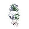
|
|---|---|
| 1 |
|
-要素
| #1: 抗体 | 分子量: 23558.154 Da / 分子数: 1 / 由来タイプ: 組換発現 / 由来: (組換発現) Mus musculus (ハツカネズミ)発現宿主:  Cricetulus griseus (モンゴルキヌゲネズミ) Cricetulus griseus (モンゴルキヌゲネズミ) |
|---|---|
| #2: 抗体 | 分子量: 23406.252 Da / 分子数: 1 / 由来タイプ: 組換発現 / 由来: (組換発現) Mus musculus (ハツカネズミ)発現宿主: Cricetulus griseus (モンゴルキヌゲネズミ) |
| #3: タンパク質 | トロンビン 分子量: 65386.113 Da / 分子数: 1 / 由来タイプ: 天然 / 由来: (天然) Homo sapiens (ヒト) / 参照: UniProt: P00734 |
| 研究の焦点であるリガンドがあるか | N |
-実験情報
-実験
| 実験 | 手法: 電子顕微鏡法 |
|---|---|
| EM実験 | 試料の集合状態: PARTICLE / 3次元再構成法: 単粒子再構成法 |
- 試料調製
試料調製
| 構成要素 | 名称: Complex of Fab fragment of POmAb with human prothrombin タイプ: COMPLEX 詳細: Fab fragment generated by proteolytic cleavage of POmAb IgG antibody in complex with open conformation of human prothrombin Entity ID: all / 由来: MULTIPLE SOURCES |
|---|---|
| 分子量 | 値: 0.12 MDa / 実験値: YES |
| 由来(天然) | 生物種: Mus musculus (ハツカネズミ) |
| 由来(組換発現) | 生物種: Cricetulus griseus (モンゴルキヌゲネズミ) |
| 緩衝液 | pH: 7.4 |
| 試料 | 濃度: 0.33 mg/ml / 包埋: NO / シャドウイング: NO / 染色: NO / 凍結: YES / 詳細: Monodisperse particles. Complex purified by SEC. |
| 試料支持 | グリッドのタイプ: Quantifoil R2/2 |
| 急速凍結 | 装置: FEI VITROBOT MARK IV / 凍結剤: ETHANE / 湿度: 95 % / 凍結前の試料温度: 277 K |
- 電子顕微鏡撮影
| 実験機器 |  モデル: Titan Krios / 画像提供: FEI Company |
|---|---|
| 顕微鏡 | モデル: FEI TITAN KRIOS |
| 電子銃 | 電子線源: FIELD EMISSION GUN / 加速電圧: 300 kV / 照射モード: FLOOD BEAM |
| 電子レンズ | モード: BRIGHT FIELDBright-field microscopy / 倍率(公称値): 59000 X / 最大 デフォーカス(公称値): 2400 nm / 最小 デフォーカス(公称値): 1000 nm / Cs: 0.01 mm / アライメント法: ZEMLIN TABLEAU |
| 試料ホルダ | 凍結剤: NITROGEN 試料ホルダーモデル: FEI TITAN KRIOS AUTOGRID HOLDER 最高温度: 84 K / 最低温度: 82 K |
| 撮影 | 電子線照射量: 62.2 e/Å2 フィルム・検出器のモデル: FEI FALCON IV (4k x 4k) |
| 電子光学装置 | 球面収差補正装置: Microscope is outfitted with a Cs image corrector with two hexapole elements. |
| 画像スキャン | 横: 4096 / 縦: 4096 |
- 解析
解析
| EMソフトウェア |
| ||||||||||||||||||||||||
|---|---|---|---|---|---|---|---|---|---|---|---|---|---|---|---|---|---|---|---|---|---|---|---|---|---|
| CTF補正 | タイプ: PHASE FLIPPING AND AMPLITUDE CORRECTION | ||||||||||||||||||||||||
| 対称性 | 点対称性: C1 (非対称) | ||||||||||||||||||||||||
| 3次元再構成 | 解像度: 3.2 Å / 解像度の算出法: FSC 0.143 CUT-OFF / 粒子像の数: 176744 / 対称性のタイプ: POINT | ||||||||||||||||||||||||
| 原子モデル構築 | 空間: REAL | ||||||||||||||||||||||||
| 拘束条件 |
|
