 Movie
Movie Controller


+ Open data

- Basic information
| Entry | Database: PDB / ID: 8ua8 | ||||||||||||||||||||||||||||||||||||||||||||||||||||||
|---|---|---|---|---|---|---|---|---|---|---|---|---|---|---|---|---|---|---|---|---|---|---|---|---|---|---|---|---|---|---|---|---|---|---|---|---|---|---|---|---|---|---|---|---|---|---|---|---|---|---|---|---|---|---|---|
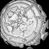
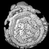
| Title | Structure of Semliki Forest virus VLP in complex with VLDLR LA2 | ||||||||||||||||||||||||||||||||||||||||||||||||||||||
 Components Components |
| ||||||||||||||||||||||||||||||||||||||||||||||||||||||
 Keywords Keywords | VIRUS LIKE PARTICLE / Semliki Forest Virus / Receptor | ||||||||||||||||||||||||||||||||||||||||||||||||||||||
| Function / homology |  Function and homology information Function and homology informationreelin receptor activity / VLDL clearance / glycoprotein transport / very-low-density lipoprotein particle receptor activity / Reelin signalling pathway / ventral spinal cord development / very-low-density lipoprotein particle binding / low-density lipoprotein particle receptor activity / very-low-density lipoprotein particle clearance / togavirin ...reelin receptor activity / VLDL clearance / glycoprotein transport / very-low-density lipoprotein particle receptor activity / Reelin signalling pathway / ventral spinal cord development / very-low-density lipoprotein particle binding / low-density lipoprotein particle receptor activity / very-low-density lipoprotein particle clearance / togavirin / reelin-mediated signaling pathway / very-low-density lipoprotein particle / positive regulation of dendrite development / T=4 icosahedral viral capsid / cargo receptor activity / dendrite morphogenesis / virion assembly / lipid transport / regulation of synapse assembly / small molecule binding / host cell endoplasmic reticulum / apolipoprotein binding / cholesterol metabolic process / clathrin-coated pit / receptor-mediated endocytosis / VLDLR internalisation and degradation / memory / calcium-dependent protein binding / nervous system development / channel activity / host cell endosome / monoatomic ion transmembrane transport / clathrin-dependent endocytosis of virus by host cell / symbiont-mediated suppression of host toll-like receptor signaling pathway / host cell Golgi apparatus / entry receptor-mediated virion attachment to host cell / signaling receptor complex / serine-type endopeptidase activity / viral translational frameshifting / lysosomal membrane / fusion of virus membrane with host endosome membrane / viral envelope / calcium ion binding / symbiont entry into host cell / host cell plasma membrane / host cell nucleus / virion membrane / structural molecule activity / glutamatergic synapse / signal transduction / proteolysis / RNA binding / membrane / plasma membrane Similarity search - Function | ||||||||||||||||||||||||||||||||||||||||||||||||||||||
| Biological species |   Semliki Forest virus Semliki Forest virus Homo sapiens (human) Homo sapiens (human) | ||||||||||||||||||||||||||||||||||||||||||||||||||||||
| Method | ELECTRON MICROSCOPY / single particle reconstruction / cryo EM / Resolution: 3.7 Å | ||||||||||||||||||||||||||||||||||||||||||||||||||||||
 Authors Authors | Abraham, J. / Yang, P. / Li, W. / Fan, X. / Pan, J. | ||||||||||||||||||||||||||||||||||||||||||||||||||||||
| Funding support |  United States, 1items United States, 1items
| ||||||||||||||||||||||||||||||||||||||||||||||||||||||
 Citation Citation | Journal: Nat Commun / Year: 2024 Title: Structural basis for VLDLR recognition by eastern equine encephalitis virus. Authors: Pan Yang / Wanyu Li / Xiaoyi Fan / Junhua Pan / Colin J Mann / Haley Varnum / Lars E Clark / Sarah A Clark / Adrian Coscia / Himanish Basu / Katherine Nabel Smith / Vesna Brusic / Jonathan Abraham /  Abstract: Eastern equine encephalitis virus (EEEV) is the most virulent alphavirus that infects humans, and many survivors develop neurological sequelae, including paralysis and intellectual disability. ...Eastern equine encephalitis virus (EEEV) is the most virulent alphavirus that infects humans, and many survivors develop neurological sequelae, including paralysis and intellectual disability. Alphavirus spike proteins comprise trimers of heterodimers of glycoproteins E2 and E1 that mediate binding to cellular receptors and fusion of virus and host cell membranes during entry. We recently identified very-low density lipoprotein receptor (VLDLR) and apolipoprotein E receptor 2 (ApoER2) as cellular receptors for EEEV and a distantly related alphavirus, Semliki Forest virus (SFV). Here, we use single-particle cryo-electron microscopy (cryo-EM) to determine structures of the EEEV and SFV spike glycoproteins bound to the VLDLR ligand-binding domain and found that EEEV and SFV interact with the same cellular receptor through divergent binding modes. Our studies suggest that the ability of LDLR-related proteins to interact with viral spike proteins through very small footprints with flexible binding modes results in a low evolutionary barrier to the acquisition of LDLR-related proteins as cellular receptors for diverse sets of viruses. | ||||||||||||||||||||||||||||||||||||||||||||||||||||||
| History |
|
- Structure visualization
| Structure viewer | Molecule: MolmilJmol/JSmol |
|---|
- Downloads & links
-Download
| PDBx/mmCIF format | 8ua8.cif.gz | 739.3 KB | Display | PDBx/mmCIF format |
|---|---|---|---|---|
| PDB format | pdb8ua8.ent.gz | 606.1 KB | Display | PDB format |
| PDBx/mmJSON format | 8ua8.json.gz | Tree view | PDBx/mmJSON format | |
| Others |  Other downloads Other downloads |
-Validation report
| Arichive directory | https://data.pdbj.org/pub/pdb/validation_reports/ua/8ua8ftp://data.pdbj.org/pub/pdb/validation_reports/ua/8ua8 | HTTPS FTP |
|---|
-Related structure data
| Related structure data |  42054MC  8ua4C  8ua9C M: map data used to model this data C: citing same article ( |
|---|---|
| Similar structure data |

-Links
 PDBj
PDBj


- Assembly
| Deposited unit | 
|
|---|---|
| 1 |
|
-Components
-Glycoprotein ... , 2 types, 8 molecules AEIMBFJN
| #1: Protein | Mass: 47489.766 Da / Num. of mol.: 4 Source method: isolated from a genetically manipulated source Source: (gene. exp.) Semliki Forest virus / Production host: Homo sapiens (human) / References: UniProt: A0A0F6PP03#2: Protein | Mass: 46330.719 Da / Num. of mol.: 4 Source method: isolated from a genetically manipulated source Source: (gene. exp.) Semliki Forest virus / Production host: Homo sapiens (human) / References: UniProt: A0A0E3T652 |
|---|
-Protein , 2 types, 8 molecules CGKODHLP
| #3: Protein | Mass: 6020.911 Da / Num. of mol.: 4 Source method: isolated from a genetically manipulated source Source: (gene. exp.) Semliki Forest virus / Production host: Homo sapiens (human) / References: UniProt: P03315#4: Protein | Mass: 16723.904 Da / Num. of mol.: 4 Source method: isolated from a genetically manipulated source Source: (gene. exp.) Semliki Forest virus / Production host: Homo sapiens (human) / References: UniProt: P03315, togavirin |
|---|
-Protein/peptide / Non-polymers , 2 types, 2 molecules R
| #5: Protein/peptide | Mass: 4028.225 Da / Num. of mol.: 1 Source method: isolated from a genetically manipulated source Source: (gene. exp.) Homo sapiens (human) / Gene: VLDLR / Production host: Homo sapiens (human) / References: UniProt: P98155 |
|---|---|
| #9: Chemical | ChemComp-CA /  Mass: 40.078 Da / Num. of mol.: 1 / Source method: obtained synthetically / Formula: Ca / Feature type: SUBJECT OF INVESTIGATION Mass: 40.078 Da / Num. of mol.: 1 / Source method: obtained synthetically / Formula: Ca / Feature type: SUBJECT OF INVESTIGATION |
-Sugars , 3 types, 14 molecules 
| #6: Polysaccharide | 2-acetamido-2-deoxy-beta-D-glucopyranose-(1-4)-2-acetamido-2-deoxy-beta-D-glucopyranose #7: Polysaccharide | beta-D-mannopyranose-(1-4)-2-acetamido-2-deoxy-beta-D-glucopyranose-(1-4)-2-acetamido-2-deoxy-beta- ...beta-D-mannopyranose-(1-4)-2-acetamido-2-deoxy-beta-D-glucopyranose-(1-4)-2-acetamido-2-deoxy-beta-D-glucopyranose #8: Sugar |  Type: D-saccharide, beta linking / Mass: 221.208 Da / Num. of mol.: 2 / Source method: obtained synthetically / Formula: C8H15NO6 / Feature type: SUBJECT OF INVESTIGATION Type: D-saccharide, beta linking / Mass: 221.208 Da / Num. of mol.: 2 / Source method: obtained synthetically / Formula: C8H15NO6 / Feature type: SUBJECT OF INVESTIGATION |
|---|
-Details
| Has ligand of interest | Y |
|---|---|
| Has protein modification | Y |
-Experimental details
-Experiment
| Experiment | Method: ELECTRON MICROSCOPY |
|---|---|
| EM experiment | Aggregation state: PARTICLE / 3D reconstruction method: single particle reconstruction |
- Sample preparation
| Component | Name: Semliki Forest virus / Type: VIRUS / Entity ID: #1-#5 / Source: RECOMBINANT |
|---|---|
| Source (natural) | Organism: Semliki Forest virus |
| Source (recombinant) | Organism: Homo sapiens (human) |
| Details of virus | Empty: NO / Enveloped: YES / Isolate: STRAIN / Type: VIRUS-LIKE PARTICLE |
| Buffer solution | pH: 7.2 |
| Specimen | Embedding applied: NO / Shadowing applied: NO / Staining applied: NO / Vitrification applied: YES |
| Vitrification | Cryogen name: ETHANE |
- Electron microscopy imaging
| Experimental equipment |  Model: Titan Krios / Image courtesy: FEI Company |
|---|---|
| Microscopy | Model: FEI TITAN KRIOS |
| Electron gun | Electron source:  FIELD EMISSION GUN / Accelerating voltage: 300 kV / Illumination mode: FLOOD BEAM FIELD EMISSION GUN / Accelerating voltage: 300 kV / Illumination mode: FLOOD BEAM |
| Electron lens | Mode: BRIGHT FIELD / Nominal defocus max: 2500 nm / Nominal defocus min: 1000 nm |
| Image recording | Electron dose: 54.9 e/Å2 / Film or detector model: GATAN K3 (6k x 4k) |
- Processing
| EM software | Name: PHENIX / Category: model refinement | ||||||||||||||||||||||||
|---|---|---|---|---|---|---|---|---|---|---|---|---|---|---|---|---|---|---|---|---|---|---|---|---|---|
| CTF correction | Type: NONE | ||||||||||||||||||||||||
| 3D reconstruction | Resolution: 3.7 Å / Resolution method: FSC 0.143 CUT-OFF / Num. of particles: 439486 / Symmetry type: POINT | ||||||||||||||||||||||||
| Refine LS restraints |
|
