 Movie
Movie Controller


+ Open data

- Basic information
| Entry | Database: PDB / ID: 8u4o | |||||||||||||||||||||||||||||||||||||||||||||
|---|---|---|---|---|---|---|---|---|---|---|---|---|---|---|---|---|---|---|---|---|---|---|---|---|---|---|---|---|---|---|---|---|---|---|---|---|---|---|---|---|---|---|---|---|---|---|
| Title | Structure of CXCL12-bound CXCR4/Gi complex | |||||||||||||||||||||||||||||||||||||||||||||
 Components Components |
| |||||||||||||||||||||||||||||||||||||||||||||
 Keywords Keywords | SIGNALING PROTEIN / GPCR / chemokine receptor / chemokine | |||||||||||||||||||||||||||||||||||||||||||||
| Function / homology |  Function and homology information Function and homology informationC-X-C motif chemokine 12 receptor activity / telencephalon cell migration / chemokine (C-X-C motif) ligand 12 signaling pathway / negative regulation of leukocyte tethering or rolling / response to ultrasound / positive regulation of macrophage migration inhibitory factor signaling pathway / regulation of actin polymerization or depolymerization / chemokine receptor binding / Specification of primordial germ cells / CXCL12-activated CXCR4 signaling pathway ...C-X-C motif chemokine 12 receptor activity / telencephalon cell migration / chemokine (C-X-C motif) ligand 12 signaling pathway / negative regulation of leukocyte tethering or rolling / response to ultrasound / positive regulation of macrophage migration inhibitory factor signaling pathway / regulation of actin polymerization or depolymerization / chemokine receptor binding / Specification of primordial germ cells / CXCL12-activated CXCR4 signaling pathway / myosin light chain binding / myelin maintenance / CXCR chemokine receptor binding / C-X-C chemokine receptor activity / positive regulation of axon extension involved in axon guidance / positive regulation of vasculature development / positive regulation of dopamine secretion / Signaling by ROBO receptors / regulation of chemotaxis / induction of positive chemotaxis / Formation of definitive endoderm / C-C chemokine receptor activity / integrin activation / negative regulation of dendritic cell apoptotic process / negative regulation of intrinsic apoptotic signaling pathway in response to DNA damage / cellular response to chemokine / chemokine-mediated signaling pathway / C-C chemokine binding / Developmental Lineage of Pancreatic Acinar Cells / positive regulation of monocyte chemotaxis / chemokine activity / Chemokine receptors bind chemokines / blood circulation / anchoring junction / dendritic cell chemotaxis / positive regulation of calcium ion import / cellular response to cytokine stimulus / cell leading edge / detection of temperature stimulus involved in sensory perception of pain / positive regulation of oligodendrocyte differentiation / animal organ regeneration / Binding and entry of HIV virion / detection of mechanical stimulus involved in sensory perception of pain / positive regulation of T cell migration / adenylate cyclase inhibitor activity / regulation of cell adhesion / Nuclear signaling by ERBB4 / positive regulation of protein localization to cell cortex / coreceptor activity / T cell migration / Adenylate cyclase inhibitory pathway / D2 dopamine receptor binding / response to prostaglandin E / adenylate cyclase regulator activity / G protein-coupled serotonin receptor binding / adenylate cyclase-inhibiting serotonin receptor signaling pathway / neurogenesis / positive regulation of endothelial cell proliferation / positive regulation of neuron differentiation / cellular response to forskolin / positive regulation of cell adhesion / axon guidance / regulation of mitotic spindle organization / adult locomotory behavior / ubiquitin binding / cell chemotaxis / Regulation of insulin secretion / growth factor activity / calcium-mediated signaling / positive regulation of cholesterol biosynthetic process / negative regulation of insulin secretion / G protein-coupled receptor activity / G protein-coupled receptor binding / defense response / response to peptide hormone / adenylate cyclase-inhibiting G protein-coupled receptor signaling pathway / brain development / adenylate cyclase-modulating G protein-coupled receptor signaling pathway / response to virus / centriolar satellite / G-protein beta/gamma-subunit complex binding / Olfactory Signaling Pathway / Activation of the phototransduction cascade / integrin binding / G beta:gamma signalling through PLC beta / Presynaptic function of Kainate receptors / Thromboxane signalling through TP receptor / intracellular calcium ion homeostasis / G protein-coupled acetylcholine receptor signaling pathway / G-protein activation / Activation of G protein gated Potassium channels / Inhibition of voltage gated Ca2+ channels via Gbeta/gamma subunits / neuron migration / Prostacyclin signalling through prostacyclin receptor / G beta:gamma signalling through CDC42 / Glucagon signaling in metabolic regulation / chemotaxis / G beta:gamma signalling through BTK / Synthesis, secretion, and inactivation of Glucagon-like Peptide-1 (GLP-1) / ADP signalling through P2Y purinoceptor 12 Similarity search - Function | |||||||||||||||||||||||||||||||||||||||||||||
| Biological species |  Homo sapiens (human) Homo sapiens (human) | |||||||||||||||||||||||||||||||||||||||||||||
| Method | ELECTRON MICROSCOPY / single particle reconstruction / cryo EM / Resolution: 3.29 Å | |||||||||||||||||||||||||||||||||||||||||||||
 Authors Authors | Saotome, K. / McGoldrick, L.L. / Franklin, M.C. | |||||||||||||||||||||||||||||||||||||||||||||
| Funding support |  United States, 1items United States, 1items
| |||||||||||||||||||||||||||||||||||||||||||||
 Citation Citation | Journal: Nat Struct Mol Biol / Year: 2025 Title: Structural insights into CXCR4 modulation and oligomerization. Authors: Kei Saotome / Luke L McGoldrick / Jo-Hao Ho / Trudy F Ramlall / Sweta Shah / Michael J Moore / Jee Hae Kim / Raymond Leidich / William C Olson / Matthew C Franklin / Abstract: Activation of the chemokine receptor CXCR4 by its chemokine ligand CXCL12 regulates diverse cellular processes. Previously reported crystal structures of CXCR4 revealed the architecture of an ...Activation of the chemokine receptor CXCR4 by its chemokine ligand CXCL12 regulates diverse cellular processes. Previously reported crystal structures of CXCR4 revealed the architecture of an inactive, homodimeric receptor. However, many structural aspects of CXCR4 remain poorly understood. Here, we use cryo-electron microscopy to investigate various modes of human CXCR4 regulation. CXCL12 activates CXCR4 by inserting its N terminus deep into the CXCR4 orthosteric pocket. The binding of US Food and Drug Administration-approved antagonist AMD3100 is stabilized by electrostatic interactions with acidic residues in the seven-transmembrane-helix bundle. A potent antibody blocker, REGN7663, binds across the extracellular face of CXCR4 and inserts its complementarity-determining region H3 loop into the orthosteric pocket. Trimeric and tetrameric structures of CXCR4 reveal modes of G-protein-coupled receptor oligomerization. We show that CXCR4 adopts distinct subunit conformations in trimeric and tetrameric assemblies, highlighting how oligomerization could allosterically regulate chemokine receptor function. #1: Journal: Biorxiv / Year: 2024Title: Structural insights into CXCR4 modulation and oligomerization Authors: Saotome, K. / McGoldrick, L.L. / Ho, J. / Ramlall, T. / Shah, S. / Moore, M.J. / Kim, J.H. / Leidich, R. / Olson, W.C. / Franklin, M.C. | |||||||||||||||||||||||||||||||||||||||||||||
| History |
|
- Structure visualization
| Structure viewer | Molecule: MolmilJmol/JSmol |
|---|
- Downloads & links
-Download
| PDBx/mmCIF format | 8u4o.cif.gz | 214.4 KB | Display | PDBx/mmCIF format |
|---|---|---|---|---|
| PDB format | pdb8u4o.ent.gz | 152.5 KB | Display | PDB format |
| PDBx/mmJSON format | 8u4o.json.gz | Tree view | PDBx/mmJSON format | |
| Others |  Other downloads Other downloads |
-Validation report
| Summary document | 8u4o_validation.pdf.gz | 1.6 MB | Display | wwPDB validaton report |
|---|---|---|---|---|
| Full document | 8u4o_full_validation.pdf.gz | 1.6 MB | Display | |
| Data in XML | 8u4o_validation.xml.gz | 37.4 KB | Display | |
| Data in CIF | 8u4o_validation.cif.gz | 54.8 KB | Display | |
| Arichive directory | https://data.pdbj.org/pub/pdb/validation_reports/u4/8u4oftp://data.pdbj.org/pub/pdb/validation_reports/u4/8u4o | HTTPS FTP |
-Related structure data
| Related structure data |  41889MC  8u4nC  8u4pC  8u4qC  8u4rC 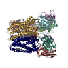 8u4sC  8u4tC M: map data used to model this data C: citing same article ( |
|---|---|
| Similar structure data |


-Links
 PDBj
PDBj


- Assembly
| Deposited unit | 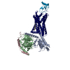
|
|---|---|
| 1 |
|
-Components
-Protein , 2 types, 2 molecules RJ
| #1: Protein | Mass: 71063.609 Da / Num. of mol.: 1 Source method: isolated from a genetically manipulated source Source: (gene. exp.) Homo sapiens (human) / Gene: CXCR4 / Production host:   Spodoptera frugiperda (fall armyworm) / References: UniProt: P61073 Spodoptera frugiperda (fall armyworm) / References: UniProt: P61073 |
|---|---|
| #5: Protein | Mass: 7978.460 Da / Num. of mol.: 1 / Fragment: UNP residues 22-89 Source method: isolated from a genetically manipulated source Source: (gene. exp.) Homo sapiens (human) / Gene: CXCL12, SDF1, SDF1A, SDF1B / Production host:  |
-Guanine nucleotide-binding protein ... , 3 types, 3 molecules ABC
| #2: Protein | Mass: 41591.312 Da / Num. of mol.: 1 Source method: isolated from a genetically manipulated source Source: (gene. exp.) Homo sapiens (human) / Gene: GNAI1 / Production host: Spodoptera frugiperda (fall armyworm) / References: UniProt: P63096 |
|---|---|
| #3: Protein | Mass: 38534.062 Da / Num. of mol.: 1 Source method: isolated from a genetically manipulated source Source: (gene. exp.) Homo sapiens (human) / Gene: GNB1 / Production host: Spodoptera frugiperda (fall armyworm) / References: UniProt: P62873 |
| #4: Protein | Mass: 7861.143 Da / Num. of mol.: 1 Source method: isolated from a genetically manipulated source Source: (gene. exp.) Homo sapiens (human) / Gene: GNG2 / Production host: Spodoptera frugiperda (fall armyworm) / References: UniProt: P59768 |
-Non-polymers , 1 types, 1 molecules 
| #6: Chemical | ChemComp-CLR /  Mass: 386.654 Da / Num. of mol.: 1 / Source method: obtained synthetically / Formula: C27H46O Mass: 386.654 Da / Num. of mol.: 1 / Source method: obtained synthetically / Formula: C27H46O |
|---|
-Details
| Has ligand of interest | N |
|---|---|
| Has protein modification | Y |
-Experimental details
-Experiment
| Experiment | Method: ELECTRON MICROSCOPY |
|---|---|
| EM experiment | Aggregation state: PARTICLE / 3D reconstruction method: single particle reconstruction |
- Sample preparation
| Component | Name: CXCL12-bound CXCR4/Gi complex / Type: COMPLEX / Entity ID: #1-#5 / Source: MULTIPLE SOURCES |
|---|---|
| Molecular weight | Experimental value: NO |
| Source (natural) | Organism: Homo sapiens (human) |
| Source (recombinant) | Organism: Spodoptera frugiperda (fall armyworm) |
| Buffer solution | pH: 7.5 |
| Specimen | Embedding applied: NO / Shadowing applied: NO / Staining applied: NO / Vitrification applied: YES |
| Vitrification | Cryogen name: ETHANE |
- Electron microscopy imaging
| Experimental equipment |  Model: Titan Krios / Image courtesy: FEI Company |
|---|---|
| Microscopy | Model: TFS KRIOS |
| Electron gun | Electron source:  FIELD EMISSION GUN / Accelerating voltage: 300 kV / Illumination mode: FLOOD BEAM FIELD EMISSION GUN / Accelerating voltage: 300 kV / Illumination mode: FLOOD BEAM |
| Electron lens | Mode: BRIGHT FIELD / Nominal defocus max: 2200 nm / Nominal defocus min: 1200 nm |
| Image recording | Electron dose: 40 e/Å2 / Film or detector model: GATAN K3 BIOQUANTUM (6k x 4k) |
- Processing
| CTF correction | Type: PHASE FLIPPING AND AMPLITUDE CORRECTION | ||||||||||||||||||||||||
|---|---|---|---|---|---|---|---|---|---|---|---|---|---|---|---|---|---|---|---|---|---|---|---|---|---|
| 3D reconstruction | Resolution: 3.29 Å / Resolution method: FSC 0.143 CUT-OFF / Num. of particles: 87963 / Symmetry type: POINT | ||||||||||||||||||||||||
| Refinement | Cross valid method: NONE Stereochemistry target values: GeoStd + Monomer Library + CDL v1.2 | ||||||||||||||||||||||||
| Displacement parameters | Biso mean: 76.5 Å2 | ||||||||||||||||||||||||
| Refine LS restraints |
|
