[English] 日本語
Yorodumi
- PDB-8u37: Crystal structure of the catalytic domain of human PKC alpha (D46... -

+
Open data


ID or keywords:

Loading...

-
Basic information

Entry
Database: PDB / ID: 8u37
TitleCrystal structure of the catalytic domain of human PKC alpha (D463N, V568I, S657E) in complex with NVP-CJL037 at 2.48-A resolution
ComponentsProtein kinase C alpha type
KeywordsTRANSFERASE/INHIBITOR / TRANSFERASE-INHIBITOR complex
Function / homology
Function and homology information


Disinhibition of SNARE formation / Response to elevated platelet cytosolic Ca2+ / positive regulation of angiotensin-activated signaling pathway / desmosome assembly / positive regulation of dense core granule biogenesis / histone H3T6 kinase activity / calcium,diacylglycerol-dependent serine/threonine kinase activity / HuR (ELAVL1) binds and stabilizes mRNA / Depolymerization of the Nuclear Lamina / Trafficking of GluR2-containing AMPA receptors ...Disinhibition of SNARE formation / Response to elevated platelet cytosolic Ca2+ / positive regulation of angiotensin-activated signaling pathway / desmosome assembly / positive regulation of dense core granule biogenesis / histone H3T6 kinase activity / calcium,diacylglycerol-dependent serine/threonine kinase activity / HuR (ELAVL1) binds and stabilizes mRNA / Depolymerization of the Nuclear Lamina / Trafficking of GluR2-containing AMPA receptors / WNT5A-dependent internalization of FZD4 / Acetylcholine regulates insulin secretion / ROBO receptors bind AKAP5 / protein kinase C / negative regulation of glial cell apoptotic process / mitotic nuclear membrane disassembly / diacylglycerol-dependent serine/threonine kinase activity / regulation of platelet aggregation / positive regulation of macrophage differentiation / positive regulation of lipopolysaccharide-mediated signaling pathway / alphav-beta3 integrin-PKCalpha complex / Calmodulin induced events / Syndecan interactions / Regulation of KIT signaling / positive regulation of adenylate cyclase-activating G protein-coupled receptor signaling pathway / positive regulation of cardiac muscle hypertrophy / RET signaling / RHO GTPases Activate NADPH Oxidases / positive regulation of cell adhesion / positive regulation of bone resorption / positive regulation of blood vessel endothelial cell migration / regulation of mRNA stability / EGFR Transactivation by Gastrin / positive regulation of endothelial cell proliferation / SHC1 events in ERBB2 signaling / positive regulation of mitotic cell cycle / response to interleukin-1 / post-translational protein modification / positive regulation of endothelial cell migration / ciliary basal body / VEGFR2 mediated cell proliferation / apoptotic signaling pathway / mitochondrial membrane / peptidyl-threonine phosphorylation / Signaling by SCF-KIT / G alpha (z) signalling events / positive regulation of angiogenesis / Inactivation, recovery and regulation of the phototransduction cascade / integrin binding / Ca2+ pathway / peptidyl-serine phosphorylation / angiogenesis / positive regulation of ERK1 and ERK2 cascade / cell adhesion / protein kinase activity / intracellular signal transduction / positive regulation of cell migration / protein phosphorylation / protein serine kinase activity / protein serine/threonine kinase activity / perinuclear region of cytoplasm / enzyme binding / endoplasmic reticulum / mitochondrion / extracellular exosome / zinc ion binding / nucleoplasm / ATP binding / plasma membrane / cytosol / cytoplasm
Similarity search - Function
Classical Protein Kinase C alpha, catalytic domain / Protein kinase C, alpha/beta/gamma types / Protein kinase, C-terminal / Protein kinase C terminal domain / Diacylglycerol/phorbol-ester binding / Phorbol esters/diacylglycerol binding domain (C1 domain) / Zinc finger phorbol-ester/DAG-type signature. / C2 domain / Protein kinase C conserved region 2 (CalB) / Zinc finger phorbol-ester/DAG-type profile. ...Classical Protein Kinase C alpha, catalytic domain / Protein kinase C, alpha/beta/gamma types / Protein kinase, C-terminal / Protein kinase C terminal domain / Diacylglycerol/phorbol-ester binding / Phorbol esters/diacylglycerol binding domain (C1 domain) / Zinc finger phorbol-ester/DAG-type signature. / C2 domain / Protein kinase C conserved region 2 (CalB) / Zinc finger phorbol-ester/DAG-type profile. / Protein kinase C conserved region 1 (C1) domains (Cysteine-rich domains) / Protein kinase C-like, phorbol ester/diacylglycerol-binding domain / C2 domain / C2 domain profile. / C1-like domain superfamily / Extension to Ser/Thr-type protein kinases / AGC-kinase, C-terminal / AGC-kinase C-terminal domain profile. / C2 domain superfamily / Serine/threonine-protein kinase, active site / Serine/Threonine protein kinases active-site signature. / Protein kinase domain / Serine/Threonine protein kinases, catalytic domain / Protein kinase, ATP binding site / Protein kinases ATP-binding region signature. / Protein kinase domain profile. / Protein kinase domain / Protein kinase-like domain superfamily
Similarity search - Domain/homology
Chem-V5U / Protein kinase C alpha type
Similarity search - Component
Biological speciesHomo sapiens (human)
MethodX-RAY DIFFRACTION / SYNCHROTRON / MOLECULAR REPLACEMENT / Resolution: 2.48 Å
AuthorsRomanowski, M.J. / Lam, J. / Visser, M.
Funding support United States, 1items
OrganizationGrant numberCountry
Other private United States
CitationJournal: J.Med.Chem. / Year: 2024
Title: Discovery of Darovasertib (NVP-LXS196), a Pan-PKC Inhibitor for the Treatment of Metastatic Uveal Melanoma.
Authors: Visser, M. / Papillon, J.P.N. / Luzzio, M. / LaMarche, M.J. / Fan, J. / Michael, W. / Wang, D. / Zhang, A. / Straub, C. / Mathieu, S. / Kato, M. / Palermo, M. / Chen, C. / Ramsey, T. / Joud, ...Authors: Visser, M. / Papillon, J.P.N. / Luzzio, M. / LaMarche, M.J. / Fan, J. / Michael, W. / Wang, D. / Zhang, A. / Straub, C. / Mathieu, S. / Kato, M. / Palermo, M. / Chen, C. / Ramsey, T. / Joud, C. / Barrett, R. / Vattay, A. / Guo, R. / Bric, A. / Chung, F. / Liang, G. / Romanowski, M.J. / Lam, J. / Thohan, S. / Atassi, F. / Wylie, A. / Cooke, V.G.
History
DepositionSep 7, 2023Deposition site: RCSB / Processing site: RCSB
Revision 1.0Jan 24, 2024Provider: repository / Type: Initial release
Revision 1.1Jan 31, 2024Group: Database references / Category: citation
Item: _citation.journal_volume / _citation.page_first / _citation.page_last

-
Structure visualization

Structure viewerMolecule:
MolmilJmol/JSmol

Downloads & links

-
Assembly

Deposited unit
A: Protein kinase C alpha type
hetero molecules


Theoretical massNumber of molelcules
Total (without water)41,2103
Polymers40,6811
Non-polymers5292
Water1267
1


  • Idetical with deposited unit
  • defined by author&software
TypeNameSymmetry operationNumber
identity operation1_555x,y,z1
Buried area110 Å2
ΔGint-7 kcal/mol
Surface area15710 Å2
MethodPISA
Unit cell
Length a, b, c (Å)107.851, 44.410, 84.789
Angle α, β, γ (deg.)90.000, 114.620, 90.000
Int Tables number5
Space group name H-MC121
Space group name HallC2y
Symmetry operation#1: x,y,z
#2: -x,y,-z
#3: x+1/2,y+1/2,z
#4: -x+1/2,y+1/2,-z

-
Components

#1: Protein Protein kinase C alpha type / PKC-A / PKC-alpha


Mass: 40681.414 Da / Num. of mol.: 1 / Fragment: UNP residues 320-672 / Mutation: D463N, V568I, S657E
Source method: isolated from a genetically manipulated source
Source: (gene. exp.) Homo sapiens (human) / Gene: PRKCA, PKCA, PRKACA / Production host: 
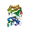
Spodoptera frugiperda (fall armyworm) / References: UniProt: P17252, protein kinase C
#2: Chemical ChemComp-V5U / (6M)-3-amino-N-{4-[(3R,4S)-4-amino-3-methoxypiperidin-1-yl]pyridin-3-yl}-6-[3-(trifluoromethoxy)pyridin-2-yl]pyrazine-2-carboxamide


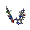
Mass: 504.465 Da / Num. of mol.: 1 / Source method: obtained synthetically / Formula: C22H23F3N8O3 / Feature type: SUBJECT OF INVESTIGATION
#3: Chemical ChemComp-MG / MAGNESIUM ION


Mass: 24.305 Da / Num. of mol.: 1 / Source method: obtained synthetically / Formula: Mg
#4: Water ChemComp-HOH / water / Water


Mass: 18.015 Da / Num. of mol.: 7 / Source method: isolated from a natural source / Formula: H2O
Has ligand of interestY

-
Experimental details

-
Experiment

ExperimentMethod: X-RAY DIFFRACTION / Number of used crystals: 1

-
Sample preparation

CrystalDensity Matthews: 2.27 Å3/Da / Density % sol: 45.79 %
Crystal growTemperature: 277 K / Method: vapor diffusion, sitting drop
Details: 0.1 M Bis-Tris, pH 5.5, 18% PEG3350, 0.2 M magnesium chloride

-
Data collection

DiffractionMean temperature: 100 K / Serial crystal experiment: N
Diffraction sourceSource: SYNCHROTRON / Site: APS / Beamline: 17-ID / Wavelength: 1 Å
DetectorType: DECTRIS PILATUS 6M / Detector: PIXEL / Date: Feb 14, 2023
RadiationProtocol: SINGLE WAVELENGTH / Monochromatic (M) / Laue (L): M / Scattering type: x-ray
Radiation wavelengthWavelength: 1 Å / Relative weight: 1
ReflectionResolution: 2.48→52.4 Å / Num. obs: 13022 / % possible obs: 98.7 % / Redundancy: 3.9 % / Biso Wilson estimate: 57.43 Å2 / Rmerge(I) obs: 0.048 / Net I/σ(I): 13.7
Reflection shellResolution: 2.48→2.62 Å / Redundancy: 3.8 % / Rmerge(I) obs: 0.372 / Mean I/σ(I) obs: 2.9 / Num. unique obs: 632 / % possible all: 98.3

-
Processing

Software
NameVersionClassification
PHENIX1.20.1_4487refinement
XDSdata reduction
XSCALEdata scaling
PHENIXphasing
RefinementMethod to determine structure: MOLECULAR REPLACEMENT
Starting model: PDB entry 3IW4
Resolution: 2.48→38.66 Å / SU ML: 0.3976 / Cross valid method: FREE R-VALUE / σ(F): 1.35 / Phase error: 35.3882
Stereochemistry target values: GeoStd + Monomer Library + CDL v1.2
RfactorNum. reflection% reflection
Rfree0.2578 633 4.88 %
Rwork0.2177 12334 -
obs0.2197 12967 98.27 %
Solvent computationShrinkage radii: 0.9 Å / VDW probe radii: 1.1 Å / Solvent model: FLAT BULK SOLVENT MODEL
Displacement parametersBiso mean: 85.34 Å2
Refinement stepCycle: LAST / Resolution: 2.48→38.66 Å
ProteinNucleic acidLigandSolventTotal
Num. atoms2592 0 37 7 2636
Refine LS restraints
Refine-IDTypeDev idealNumber
X-RAY DIFFRACTIONf_bond_d0.00772725
X-RAY DIFFRACTIONf_angle_d1.41153690
X-RAY DIFFRACTIONf_chiral_restr0.0749387
X-RAY DIFFRACTIONf_plane_restr0.0124474
X-RAY DIFFRACTIONf_dihedral_angle_d7.3984360
LS refinement shell
Resolution (Å)Rfactor RfreeNum. reflection RfreeRfactor RworkNum. reflection RworkRefine-ID% reflection obs (%)
2.48-2.670.4371400.35962423X-RAY DIFFRACTION97.9
2.67-2.940.42671170.32252472X-RAY DIFFRACTION98.55
2.94-3.370.34121330.27082424X-RAY DIFFRACTION98.57
3.37-4.240.20971250.20192485X-RAY DIFFRACTION98.6
4.24-38.660.20761180.17442530X-RAY DIFFRACTION97.75
Refinement TLS params.Method: refined / Origin x: 24.1208186709 Å / Origin y: -0.501735404402 Å / Origin z: 16.4102424511 Å
111213212223313233
T0.481563701162 Å2-0.0287485644631 Å2-0.00602833803717 Å2-0.450903016907 Å20.0203660198482 Å2--0.443671869015 Å2
L0.454283390679 °20.481428272173 °2-0.212438659274 °2-1.60998899491 °2-0.508412707694 °2--1.47359686243 °2
S-0.0501364819541 Å °-0.094150206624 Å °0.0535188932953 Å °0.376115106152 Å °-0.0273064696291 Å °0.0437405713583 Å °-0.112836079716 Å °0.0387218661636 Å °-0.000255149034118 Å °
Refinement TLS groupSelection details: all

+
About Yorodumi

-
News

-
Feb 9, 2022. New format data for meta-information of EMDB entries

New format data for meta-information of EMDB entries

  • Version 3 of the EMDB header file is now the official format.
  • The previous official version 1.9 will be removed from the archive.

Related info.:EMDB header

External links:wwPDB to switch to version 3 of the EMDB data model

-
Aug 12, 2020. Covid-19 info

Covid-19 info

URL: https://pdbj.org/emnavi/covid19.php

New page: Covid-19 featured information page in EM Navigator.

Related info.:Covid-19 info / Mar 5, 2020. Novel coronavirus structure data

+
Mar 5, 2020. Novel coronavirus structure data

Novel coronavirus structure data

Related info.:Yorodumi Speices / Aug 12, 2020. Covid-19 info

External links:COVID-19 featured content - PDBj / Molecule of the Month (242):Coronavirus Proteases

+
Jan 31, 2019. EMDB accession codes are about to change! (news from PDBe EMDB page)

EMDB accession codes are about to change! (news from PDBe EMDB page)

  • The allocation of 4 digits for EMDB accession codes will soon come to an end. Whilst these codes will remain in use, new EMDB accession codes will include an additional digit and will expand incrementally as the available range of codes is exhausted. The current 4-digit format prefixed with “EMD-” (i.e. EMD-XXXX) will advance to a 5-digit format (i.e. EMD-XXXXX), and so on. It is currently estimated that the 4-digit codes will be depleted around Spring 2019, at which point the 5-digit format will come into force.
  • The EM Navigator/Yorodumi systems omit the EMD- prefix.

Related info.:Q: What is EMD? / ID/Accession-code notation in Yorodumi/EM Navigator

External links:EMDB Accession Codes are Changing Soon! / Contact to PDBj

+
Jul 12, 2017. Major update of PDB

Major update of PDB

  • wwPDB released updated PDB data conforming to the new PDBx/mmCIF dictionary.
  • This is a major update changing the version number from 4 to 5, and with Remediation, in which all the entries are updated.
  • In this update, many items about electron microscopy experimental information are reorganized (e.g. em_software).
  • Now, EM Navigator and Yorodumi are based on the updated data.

External links:wwPDB Remediation / Enriched Model Files Conforming to OneDep Data Standards Now Available in the PDB FTP Archive

-
Yorodumi

Thousand views of thousand structures

  • Yorodumi is a browser for structure data from EMDB, PDB, SASBDB, etc.
  • This page is also the successor to EM Navigator detail page, and also detail information page/front-end page for Omokage search.
  • The word "yorodu" (or yorozu) is an old Japanese word meaning "ten thousand". "mi" (miru) is to see.

Related info.:EMDB / PDB / SASBDB / Comparison of 3 databanks / Yorodumi Search / Aug 31, 2016. New EM Navigator & Yorodumi / Yorodumi Papers / Jmol/JSmol / Function and homology information / Changes in new EM Navigator and Yorodumi

Read more