 Movie
Movie Controller

[English] 日本語
 Yorodumi
- PDB-8u10: In situ cryo-EM structure of bacteriophage P22 gp1:gp4:gp5:gp10:g... -
+ Open data

- Basic information
| Entry | Database: PDB / ID: 8u10 | ||||||
|---|---|---|---|---|---|---|---|
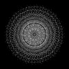
| Title | In situ cryo-EM structure of bacteriophage P22 gp1:gp4:gp5:gp10:gp9 N-term complex in conformation 1 at 3.2A resolution | ||||||
 Components Components |
| ||||||
 Keywords Keywords | VIRAL PROTEIN / phage / bacteriophage / tail spike protein / TSP / gene product 9 (gp9) / Packaged DNA stabilization protein / gene product 10 (gp10) / STRUCTURAL PROTEIN / head-to-tail protein / gene product 4 (gp4) / Tail hub protein / gene product (1) / Portal protein / Coat protein / Major capsid protein / gene product 5 (gp5) | ||||||
| Function / homology |  Function and homology information Function and homology informationviral DNA genome packaging, headful / symbiont entry into host cell via disruption of host cell wall peptidoglycan / endo-1,3-alpha-L-rhamnosidase activity / symbiont entry into host cell via disruption of host cell envelope lipopolysaccharide / viral procapsid / viral portal complex / viral procapsid maturation / symbiont genome ejection through host cell envelope, short tail mechanism / viral DNA genome packaging / virus tail, fiber ...viral DNA genome packaging, headful / symbiont entry into host cell via disruption of host cell wall peptidoglycan / endo-1,3-alpha-L-rhamnosidase activity / symbiont entry into host cell via disruption of host cell envelope lipopolysaccharide / viral procapsid / viral portal complex / viral procapsid maturation / symbiont genome ejection through host cell envelope, short tail mechanism / viral DNA genome packaging / virus tail, fiber / symbiont entry into host cell via disruption of host cell envelope / virus tail / symbiont entry into host / T=7 icosahedral viral capsid / Hydrolases; Glycosylases; Glycosidases, i.e. enzymes that hydrolyse O- and S-glycosyl compounds / adhesion receptor-mediated virion attachment to host cell / virion assembly / virion component / viral capsid / lyase activity / hydrolase activity / virion attachment to host cell / identical protein binding Similarity search - Function | ||||||
| Biological species |  Salmonella phage P22 (virus) Salmonella phage P22 (virus) | ||||||
| Method | ELECTRON MICROSCOPY / single particle reconstruction / cryo EM / Resolution: 3.2 Å | ||||||
 Authors Authors | Iglesias, S. / Feng-Hou, C. / Cingolani, G. | ||||||
| Funding support |  United States, 1items United States, 1items
| ||||||
 Citation Citation | Journal: J Mol Biol / Year: 2023 Title: Molecular Architecture of Salmonella Typhimurium Virus P22 Genome Ejection Machinery. Authors: Stephano M Iglesias / Ravi K Lokareddy / Ruoyu Yang / Fenglin Li / Daniel P Yeggoni / Chun-Feng David Hou / Makayla N Leroux / Juliana R Cortines / Justin C Leavitt / Mary Bird / Sherwood R ...Authors: Stephano M Iglesias / Ravi K Lokareddy / Ruoyu Yang / Fenglin Li / Daniel P Yeggoni / Chun-Feng David Hou / Makayla N Leroux / Juliana R Cortines / Justin C Leavitt / Mary Bird / Sherwood R Casjens / Simon White / Carolyn M Teschke / Gino Cingolani /  Abstract: Bacteriophage P22 is a prototypical member of the Podoviridae superfamily. Since its discovery in 1952, P22 has become a paradigm for phage transduction and a model for icosahedral viral capsid ...Bacteriophage P22 is a prototypical member of the Podoviridae superfamily. Since its discovery in 1952, P22 has become a paradigm for phage transduction and a model for icosahedral viral capsid assembly. Here, we describe the complete architecture of the P22 tail apparatus (gp1, gp4, gp10, gp9, and gp26) and the potential location and organization of P22 ejection proteins (gp7, gp20, and gp16), determined using cryo-EM localized reconstruction, genetic knockouts, and biochemical analysis. We found that the tail apparatus exists in two equivalent conformations, rotated by ∼6° relative to the capsid. Portal protomers make unique contacts with coat subunits in both conformations, explaining the 12:5 symmetry mismatch. The tail assembles around the hexameric tail hub (gp10), which folds into an interrupted β-propeller characterized by an apical insertion domain. The tail hub connects proximally to the dodecameric portal protein and head-to-tail adapter (gp4), distally to the trimeric tail needle (gp26), and laterally to six trimeric tailspikes (gp9) that attach asymmetrically to gp10 insertion domain. Cryo-EM analysis of P22 mutants lacking the ejection proteins gp7 or gp20 and biochemical analysis of purified recombinant proteins suggest that gp7 and gp20 form a molecular complex associated with the tail apparatus via the portal protein barrel. We identified a putative signal transduction pathway from the tailspike to the tail needle, mediated by three flexible loops in the tail hub, that explains how lipopolysaccharide (LPS) is sufficient to trigger the ejection of the P22 DNA in vitro. | ||||||
| History |
|
- Structure visualization
| Structure viewer | Molecule: MolmilJmol/JSmol |
|---|
- Downloads & links
-Download
| PDBx/mmCIF format | 8u10.cif.gz | 3.5 MB | Display | PDBx/mmCIF format |
|---|---|---|---|---|
| PDB format | pdb8u10.ent.gz | Display | PDB format | |
| PDBx/mmJSON format | 8u10.json.gz | Tree view | PDBx/mmJSON format | |
| Others |  Other downloads Other downloads |
-Validation report
| Arichive directory | https://data.pdbj.org/pub/pdb/validation_reports/u1/8u10ftp://data.pdbj.org/pub/pdb/validation_reports/u1/8u10 | HTTPS FTP |
|---|
-Related structure data
| Related structure data | 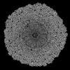 41791MC  8tvrC 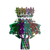 8tvuC  8u11C 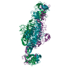 8u1oC M: map data used to model this data C: citing same article ( |
|---|---|
| Similar structure data |

-Links
 PDBj
PDBj
- Assembly
| Deposited unit | 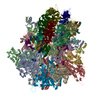
|
|---|---|
| 1 |
|
-Components
| #1: Protein | Mass: 82829.375 Da / Num. of mol.: 12 / Source method: isolated from a natural source / Source: (natural) Salmonella phage P22 (virus) / References: UniProt: P26744#2: Protein | Mass: 46795.613 Da / Num. of mol.: 10 / Source method: isolated from a natural source / Source: (natural) Salmonella phage P22 (virus) / References: UniProt: P26747#3: Protein | Mass: 52512.020 Da / Num. of mol.: 6 / Source method: isolated from a natural source / Source: (natural) Salmonella phage P22 (virus) / References: UniProt: P26749#4: Protein | Mass: 71923.523 Da / Num. of mol.: 18 / Source method: isolated from a natural source / Source: (natural) Salmonella phage P22 (virus) / References: UniProt: P12528#5: Protein | Mass: 18044.959 Da / Num. of mol.: 12 / Source method: isolated from a natural source / Source: (natural) Salmonella phage P22 (virus) / References: UniProt: P26746 |
|---|
-Experimental details
-Experiment
| Experiment | Method: ELECTRON MICROSCOPY |
|---|---|
| EM experiment | Aggregation state: PARTICLE / 3D reconstruction method: single particle reconstruction |
- Sample preparation
| Component | Name: Salmonella phage P22 / Type: VIRUS / Entity ID: all / Source: NATURAL |
|---|---|
| Source (natural) | Organism: Salmonella phage P22 (virus) |
| Details of virus | Empty: NO / Enveloped: NO / Isolate: OTHER / Type: VIRION |
| Natural host | Organism: Salmonella enterica |
| Buffer solution | pH: 7.5 |
| Specimen | Embedding applied: NO / Shadowing applied: NO / Staining applied: NO / Vitrification applied: YES |
| Vitrification | Instrument: FEI VITROBOT MARK IV / Cryogen name: ETHANE / Humidity: 100 % / Chamber temperature: 277.15 K |
- Electron microscopy imaging
| Experimental equipment |  Model: Titan Krios / Image courtesy: FEI Company |
|---|---|
| Microscopy | Model: TFS KRIOS |
| Electron gun | Electron source:  FIELD EMISSION GUN / Accelerating voltage: 300 kV / Illumination mode: OTHER FIELD EMISSION GUN / Accelerating voltage: 300 kV / Illumination mode: OTHER |
| Electron lens | Mode: OTHER / Nominal magnification: 29000 X / Calibrated magnification: 29000 X / Nominal defocus max: 2100 nm / Nominal defocus min: 800 nm / Calibrated defocus min: 800 nm / Calibrated defocus max: 2100 nm / Cs: 2.7 mm / C2 aperture diameter: 100 µm |
| Specimen holder | Cryogen: NITROGEN / Specimen holder model: FEI TITAN KRIOS AUTOGRID HOLDER |
| Image recording | Electron dose: 1.08 e/Å2 / Film or detector model: GATAN K3 (6k x 4k) |
| Image scans | Width: 11520 / Height: 8184 |
- Processing
| EM software |
| ||||||||||||||||||||||||
|---|---|---|---|---|---|---|---|---|---|---|---|---|---|---|---|---|---|---|---|---|---|---|---|---|---|
| CTF correction | Type: PHASE FLIPPING AND AMPLITUDE CORRECTION | ||||||||||||||||||||||||
| Symmetry | Point symmetry: C1 (asymmetric) | ||||||||||||||||||||||||
| 3D reconstruction | Resolution: 3.2 Å / Resolution method: FSC 0.143 CUT-OFF / Num. of particles: 18053 / Num. of class averages: 1 / Symmetry type: POINT | ||||||||||||||||||||||||
| Atomic model building | Protocol: FLEXIBLE FIT / Space: REAL | ||||||||||||||||||||||||
| Refinement | Cross valid method: NONE Stereochemistry target values: GeoStd + Monomer Library + CDL v1.2 | ||||||||||||||||||||||||
| Displacement parameters | Biso mean: 67.93 Å2 | ||||||||||||||||||||||||
| Refine LS restraints |
|
