 Movie
Movie Controller

[English] 日本語
 Yorodumi
- EMDB-41651: In situ cryo-EM structure of bacteriophage P22 portal protein: he... -
+ Open data

- Basic information
| Entry |  | |||||||||
|---|---|---|---|---|---|---|---|---|---|---|
| Title | In situ cryo-EM structure of bacteriophage P22 portal protein: head-to-tail protein complex at 3.0A resolution | |||||||||
 Map data Map data | ||||||||||
 Sample Sample |
| |||||||||
 Keywords Keywords | phage / bacteriophage / portal protein / head-to-tail protein / gene product 1 (gp1) / gene product 4 (gp4 / STRUCTURAL PROTEIN / VIRAL PROTEIN | |||||||||
| Function / homology |  Function and homology information Function and homology informationviral DNA genome packaging, headful / symbiont entry into host cell via disruption of host cell wall peptidoglycan / viral portal complex / symbiont genome ejection through host cell envelope, short tail mechanism / viral DNA genome packaging / symbiont entry into host cell via disruption of host cell envelope / virus tail / virion assembly / hydrolase activity Similarity search - Function | |||||||||
| Biological species |  Salmonella phage P22 (virus) Salmonella phage P22 (virus) | |||||||||
| Method | single particle reconstruction / cryo EM / Resolution: 3.0 Å | |||||||||
 Authors Authors | Iglesias SM / Cingolani G / Feng-Hou C | |||||||||
| Funding support |  United States, 1 items United States, 1 items
| |||||||||
 Citation Citation | Journal: J Mol Biol / Year: 2023 Title: Molecular Architecture of Salmonella Typhimurium Virus P22 Genome Ejection Machinery. Authors: Stephano M Iglesias / Ravi K Lokareddy / Ruoyu Yang / Fenglin Li / Daniel P Yeggoni / Chun-Feng David Hou / Makayla N Leroux / Juliana R Cortines / Justin C Leavitt / Mary Bird / Sherwood R ...Authors: Stephano M Iglesias / Ravi K Lokareddy / Ruoyu Yang / Fenglin Li / Daniel P Yeggoni / Chun-Feng David Hou / Makayla N Leroux / Juliana R Cortines / Justin C Leavitt / Mary Bird / Sherwood R Casjens / Simon White / Carolyn M Teschke / Gino Cingolani /  Abstract: Bacteriophage P22 is a prototypical member of the Podoviridae superfamily. Since its discovery in 1952, P22 has become a paradigm for phage transduction and a model for icosahedral viral capsid ...Bacteriophage P22 is a prototypical member of the Podoviridae superfamily. Since its discovery in 1952, P22 has become a paradigm for phage transduction and a model for icosahedral viral capsid assembly. Here, we describe the complete architecture of the P22 tail apparatus (gp1, gp4, gp10, gp9, and gp26) and the potential location and organization of P22 ejection proteins (gp7, gp20, and gp16), determined using cryo-EM localized reconstruction, genetic knockouts, and biochemical analysis. We found that the tail apparatus exists in two equivalent conformations, rotated by ∼6° relative to the capsid. Portal protomers make unique contacts with coat subunits in both conformations, explaining the 12:5 symmetry mismatch. The tail assembles around the hexameric tail hub (gp10), which folds into an interrupted β-propeller characterized by an apical insertion domain. The tail hub connects proximally to the dodecameric portal protein and head-to-tail adapter (gp4), distally to the trimeric tail needle (gp26), and laterally to six trimeric tailspikes (gp9) that attach asymmetrically to gp10 insertion domain. Cryo-EM analysis of P22 mutants lacking the ejection proteins gp7 or gp20 and biochemical analysis of purified recombinant proteins suggest that gp7 and gp20 form a molecular complex associated with the tail apparatus via the portal protein barrel. We identified a putative signal transduction pathway from the tailspike to the tail needle, mediated by three flexible loops in the tail hub, that explains how lipopolysaccharide (LPS) is sufficient to trigger the ejection of the P22 DNA in vitro. | |||||||||
| History |
|
- Structure visualization
| Supplemental images |
|---|

- Downloads & links
-EMDB archive
| Map data | emd_41651.map.gz | 191.4 MB | EMDB map data format | |
|---|---|---|---|---|
| Header (meta data) | emd-41651-v30.xmlemd-41651.xml | 18.6 KB 18.6 KB | Display Display | EMDB header |
| FSC (resolution estimation) | emd_41651_fsc.xml | 15.5 KB | Display | FSC data file |
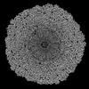
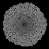
| Images | 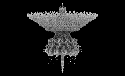 emd_41651.png emd_41651.png | 41.5 KB | ||
| Masks | emd_41651_msk_1.map | 325 MB | Mask map | |
| Filedesc metadata | emd-41651.cif.gz | 6.2 KB | ||
| Others | emd_41651_half_map_1.map.gzemd_41651_half_map_2.map.gz | 26.1 MB 26.1 MB | ||
| Archive directory |  http://ftp.pdbj.org/pub/emdb/structures/EMD-41651ftp://ftp.pdbj.org/pub/emdb/structures/EMD-41651 http://ftp.pdbj.org/pub/emdb/structures/EMD-41651ftp://ftp.pdbj.org/pub/emdb/structures/EMD-41651 | HTTPS FTP |
-Related structure data
| Related structure data |  8tvuMC  8tvrC  8u10C  8u11C 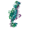 8u1oC M: atomic model generated by this map C: citing same article ( |
|---|---|
| Similar structure data |

-Links
| EMDB pages | EMDB (EBI/PDBe) / EMDataResource |
|---|
-Map
| File | Download / File: emd_41651.map.gz / Format: CCP4 / Size: 325 MB / Type: IMAGE STORED AS FLOATING POINT NUMBER (4 BYTES) | ||||||||||||||||||||||||||||||||||||
|---|---|---|---|---|---|---|---|---|---|---|---|---|---|---|---|---|---|---|---|---|---|---|---|---|---|---|---|---|---|---|---|---|---|---|---|---|---|
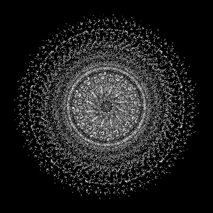
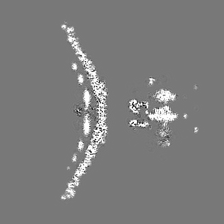
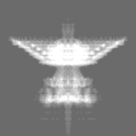
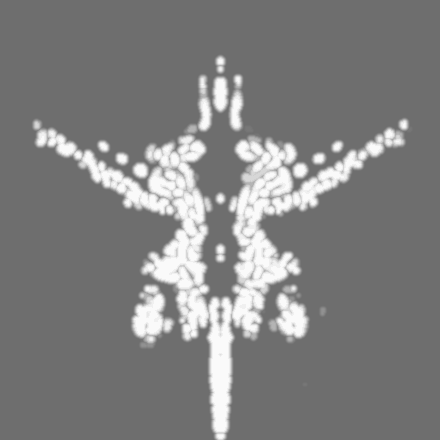
| Projections & slices | Image control
Images are generated by Spider. | ||||||||||||||||||||||||||||||||||||
| Voxel size | X=Y=Z: 1.16582 Å | ||||||||||||||||||||||||||||||||||||
| Density |
| ||||||||||||||||||||||||||||||||||||
| Symmetry | Space group: 1 | ||||||||||||||||||||||||||||||||||||
| Details | EMDB XML:
|
 X (Sec.)
 Y (Row.)
 Z (Col.)


-Supplemental data
-Mask #1
| File | emd_41651_msk_1.map | ||||||||||||
|---|---|---|---|---|---|---|---|---|---|---|---|---|---|
| Projections & Slices |
| ||||||||||||
| Density Histograms |


-Half map: #2
| File | emd_41651_half_map_1.map | ||||||||||||
|---|---|---|---|---|---|---|---|---|---|---|---|---|---|
| Projections & Slices |
| ||||||||||||
| Density Histograms |


-Half map: #1
| File | emd_41651_half_map_2.map | ||||||||||||
|---|---|---|---|---|---|---|---|---|---|---|---|---|---|
| Projections & Slices |
| ||||||||||||
| Density Histograms |


- Sample components
-Entire : Salmonella phage P22
| Entire | Name: Salmonella phage P22 (virus) |
|---|---|
| Components |
|
-Supramolecule #1: Salmonella phage P22
| Supramolecule | Name: Salmonella phage P22 / type: virus / ID: 1 / Parent: 0 / Macromolecule list: all / NCBI-ID: 2908168 / Sci species name: Salmonella phage P22 / Virus type: VIRION / Virus isolate: OTHER / Virus enveloped: No / Virus empty: No |
|---|---|
| Host (natural) | Organism:  Salmonella enterica (bacteria) Salmonella enterica (bacteria) |
-Macromolecule #1: Portal protein
| Macromolecule | Name: Portal protein / type: protein_or_peptide / ID: 1 / Number of copies: 12 / Enantiomer: LEVO |
|---|---|
| Source (natural) | Organism: Salmonella phage P22 (virus) |
| Molecular weight | Theoretical: 82.829375 KDa |
| Sequence | String: MADNENRLES ILSRFDADWT ASDEARREAK NDLFFSRVSQ WDDWLSQYTT LQYRGQFDVV RPVVRKLVSE MRQNPIDVLY RPKDGARPD AADVLMGMYR TDMRHNTAKI AVNIAVREQI EAGVGAWRLV TDYEDQSPTS NNQVIRREPI HSACSHVIWD S NSKLMDKS ...String: MADNENRLES ILSRFDADWT ASDEARREAK NDLFFSRVSQ WDDWLSQYTT LQYRGQFDVV RPVVRKLVSE MRQNPIDVLY RPKDGARPD AADVLMGMYR TDMRHNTAKI AVNIAVREQI EAGVGAWRLV TDYEDQSPTS NNQVIRREPI HSACSHVIWD S NSKLMDKS DARHCTVIHS MSQNGWEDFA EKYDLDADDI PSFQNPNDWV FPWLTQDTIQ IAEFYEVVEK KETAFIYQDP VT GEPVSYF KRDIKDVIDD LADSGFIKIA ERQIKRRRVY KSIITCTAVL KDKQLIAGEH IPIVPVFGEW GFVEDKEVYE GVV RLTKDG QRLRNMIMSF NADIVARTPK KKPFFWPEQI AGFEHMYDGN DDYPYYLLNR TDENSGDLPT QPLAYYENPE VPQA NAYML EAATSAVKEV ATLGVDTEAV NGGQVAFDTV NQLNMRADLE TYVFQDNLAT AMRRDGEIYQ SIVNDIYDVP RNVTI TLED GSEKDVQLMA EVVDLATGEK QVLNDIRGRY ECYTDVGPSF QSMKQQNRAE ILELLGKTPQ GTPEYQLLLL QYFTLL DGK GVEMMRDYAN KQLIQMGVKK PETPEEQQWL VEAQQAKQGQ QDPAMVQAQG VLLQGQAELA KAQNQTLSLQ IDAAKVE AQ NQLNAARIAE IFNNMDLSKQ SEFREFLKTV ASFQQDRSED ARANAELLLK GDEQTHKQRM DIANILQSQR QNQPSGSV A ETPQ UniProtKB: Portal protein |
-Macromolecule #2: Peptidoglycan hydrolase gp4
| Macromolecule | Name: Peptidoglycan hydrolase gp4 / type: protein_or_peptide / ID: 2 / Number of copies: 12 / Enantiomer: LEVO |
|---|---|
| Source (natural) | Organism: Salmonella phage P22 (virus) |
| Molecular weight | Theoretical: 18.044959 KDa |
| Sequence | String: MQIKTKGDLV RAALRKLGVA SDATLTDVEP QSMQDAVDDL EAMMAEWYQD GKGIITGYVF SDDENPPAEG DDHGLRSSAV SAVFHNLAC RIAPDYALEA TAKIIATAKY GKELLYKQTA ISRAKRAPYP SRMPTGSGNS FANLNEWHYF PGEQNADSTT P HDEGNG UniProtKB: Head-to-tail adapter gp4 |
-Experimental details
-Structure determination
| Method | cryo EM |
|---|---|
 Processing Processing | single particle reconstruction |
| Aggregation state | particle |
-Sample preparation
| Buffer | pH: 7.5 |
|---|---|
| Grid | Model: Quantifoil R2/1 / Material: COPPER / Mesh: 300 / Support film - Material: CARBON / Support film - topology: HOLEY / Pretreatment - Type: GLOW DISCHARGE / Pretreatment - Time: 15 sec. / Pretreatment - Atmosphere: OTHER / Pretreatment - Pressure: 0.015 kPa |
| Vitrification | Cryogen name: ETHANE / Chamber humidity: 100 % / Chamber temperature: 277.15 K / Instrument: FEI VITROBOT MARK IV |
- Electron microscopy
| Microscope | TFS KRIOS |
|---|---|
| Image recording | Film or detector model: GATAN K3 (6k x 4k) / Digitization - Dimensions - Width: 11520 pixel / Digitization - Dimensions - Height: 8184 pixel / Average electron dose: 1.08 e/Å2 |
| Electron beam | Acceleration voltage: 300 kV / Electron source:  FIELD EMISSION GUN FIELD EMISSION GUN |
| Electron optics | C2 aperture diameter: 100.0 µm / Calibrated defocus max: 2.1 µm / Calibrated defocus min: 0.8 µm / Calibrated magnification: 29000 / Illumination mode: OTHER / Imaging mode: OTHER / Cs: 2.7 mm / Nominal defocus max: 2.1 µm / Nominal defocus min: 0.8 µm / Nominal magnification: 29000 |
| Sample stage | Specimen holder model: FEI TITAN KRIOS AUTOGRID HOLDER / Cooling holder cryogen: NITROGEN |
| Experimental equipment |  Model: Titan Krios / Image courtesy: FEI Company |

