 Movie
Movie Controller

[English] 日本語
 Yorodumi
- PDB-8u0g: Full-length dimer of DNA-Damage Response Protein C from Deinococc... -
+ Open data

- Basic information
| Entry | Database: PDB / ID: 8u0g | ||||||
|---|---|---|---|---|---|---|---|
| Title | Full-length dimer of DNA-Damage Response Protein C from Deinococcus radiodurans - Crystal form xMJ7124 | ||||||
 Components Components | DNA damage response protein C | ||||||
 Keywords Keywords | DNA BINDING PROTEIN / DNA Repair / Radioresistance | ||||||
| Function / homology | cellular response to desiccation / nucleoid / cellular response to gamma radiation / DNA repair / DNA damage response / DNA damage response protein C Function and homology information Function and homology information | ||||||
| Biological species |  Deinococcus radiodurans R1 = ATCC 13939 = DSM 20539 (radioresistant) Deinococcus radiodurans R1 = ATCC 13939 = DSM 20539 (radioresistant) | ||||||
| Method |  X-RAY DIFFRACTION / MOLECULAR REPLACEMENT / Resolution: 4.28 Å X-RAY DIFFRACTION / MOLECULAR REPLACEMENT / Resolution: 4.28 Å | ||||||
 Authors Authors | Szabla, R. / Li, M.C. / Junop, M.S. | ||||||
| Funding support |  Canada, 1items Canada, 1items
| ||||||
 Citation Citation | Journal: Nucleic Acids Res. / Year: 2024 Title: DdrC, a unique DNA repair factor from D. radiodurans, senses and stabilizes DNA breaks through a novel lesion-recognition mechanism. Authors: Szabla, R. / Li, M. / Warner, V. / Song, Y. / Junop, M. | ||||||
| History |
|
- Structure visualization
| Structure viewer | Molecule: MolmilJmol/JSmol |
|---|
- Downloads & links
-Download
| PDBx/mmCIF format | 8u0g.cif.gz | 172.2 KB | Display | PDBx/mmCIF format |
|---|---|---|---|---|
| PDB format | pdb8u0g.ent.gz | 136.7 KB | Display | PDB format |
| PDBx/mmJSON format | 8u0g.json.gz | Tree view | PDBx/mmJSON format | |
| Others |  Other downloads Other downloads |
-Validation report
| Arichive directory | https://data.pdbj.org/pub/pdb/validation_reports/u0/8u0gftp://data.pdbj.org/pub/pdb/validation_reports/u0/8u0g | HTTPS FTP |
|---|
-Related structure data
| Related structure data |  7udiC 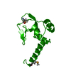 8u1jC C: citing same article ( |
|---|---|
| Similar structure data | |
| Experimental dataset #1 | Data reference: 10.5281/zenodo.8302395 / Data set type: diffraction image data |
-Links
 PDBj
PDBj- Assembly
| Deposited unit | 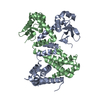
| ||||||||||||
|---|---|---|---|---|---|---|---|---|---|---|---|---|---|
| 1 |
| ||||||||||||
| Unit cell |
|
-Components
| #1: Protein | Mass: 25241.369 Da / Num. of mol.: 2 Source method: isolated from a genetically manipulated source Source: (gene. exp.) Deinococcus radiodurans R1 = ATCC 13939 = DSM 20539 (radioresistant)Strain: R1 / Gene: ddrC / Plasmid: pMJ5741 Details (production host): WT DdrC with N-term His-tag and TEV site in pDEST-527 backbone Production host: |
|---|
-Experimental details
-Experiment
| Experiment | Method: X-RAY DIFFRACTION / Number of used crystals: 1 |
|---|
- Sample preparation
| Crystal | Density Matthews: 3.48 Å3/Da / Density % sol: 64.6 % Description: Hexagonal bipyramid ~100um in width, depth and height |
|---|---|
| Crystal grow | Temperature: 293 K / Method: vapor diffusion, hanging drop Details: 1.5 ul of protein solution was mixed with 1.5 uL of crystallization solution and hung upside-down in a sealed chamber containing 1mL of well solution Protein solution: 140uM DdrC, 200mM ...Details: 1.5 ul of protein solution was mixed with 1.5 uL of crystallization solution and hung upside-down in a sealed chamber containing 1mL of well solution Protein solution: 140uM DdrC, 200mM Sodium sulfate, 1mM Magnesium chloride, 20mM Sodium citrate / Citric acid, pH 6.5 Crystallization solution (Wizard Classics 1 #22) 10% (v/v) 2-propanol 100mM Tris-base/HCl, pH 8.5 Well solution: 1.5M Ammonium sulfate PH range: 6.0 - 8.5 / Temp details: Temperature-controlled incubator at 20C |
-Data collection
| Diffraction | Mean temperature: 100 K / Ambient temp details: Nitrogen cryo-stream / Serial crystal experiment: N |
|---|---|
| Diffraction source | Source: ROTATING ANODE / Type: RIGAKU MICROMAX-007 HF / Wavelength: 1.54178 Å |
| Detector | Type: RIGAKU SATURN 944+ / Detector: CCD / Date: Sep 23, 2022 |
| Radiation | Protocol: SINGLE WAVELENGTH / Monochromatic (M) / Laue (L): M / Scattering type: x-ray |
| Radiation wavelength | Wavelength: 1.54178 Å / Relative weight: 1 |
| Reflection | Resolution: 4.277→96.163 Å / Num. obs: 5236 / % possible obs: 100 % / Redundancy: 8.9 % / Biso Wilson estimate: 170.43 Å2 / CC1/2: 0.998 / Rmerge(I) obs: 0.179 / Rpim(I) all: 0.064 / Net I/σ(I): 9.9 |
| Reflection shell | Resolution: 4.277→4.351 Å / Redundancy: 9.1 % / Rmerge(I) obs: 0.937 / Mean I/σ(I) obs: 2.9 / Num. unique obs: 242 / CC1/2: 0.889 / Rpim(I) all: 0.344 / % possible all: 100 |
- Processing
| Software |
| ||||||||||||||||||||||||
|---|---|---|---|---|---|---|---|---|---|---|---|---|---|---|---|---|---|---|---|---|---|---|---|---|---|
| Refinement | Method to determine structure: MOLECULAR REPLACEMENT / Resolution: 4.28→69.81 Å / SU ML: 0.4185 / Cross valid method: FREE R-VALUE / σ(F): 1.34 / Phase error: 29.0992 Stereochemistry target values: GeoStd + Monomer Library + CDL v1.2
| ||||||||||||||||||||||||
| Solvent computation | Shrinkage radii: 0.9 Å / VDW probe radii: 1.11 Å / Solvent model: FLAT BULK SOLVENT MODEL | ||||||||||||||||||||||||
| Displacement parameters | Biso mean: 215.44 Å2 | ||||||||||||||||||||||||
| Refinement step | Cycle: LAST / Resolution: 4.28→69.81 Å
| ||||||||||||||||||||||||
| Refine LS restraints |
| ||||||||||||||||||||||||
| LS refinement shell |
|
