 Movie
Movie Controller

[English] 日本語
 Yorodumi
- PDB-7udi: Full-length dimer of DNA-Damage Response Protein C from Deinococc... -
+ Open data

- Basic information
| Entry | Database: PDB / ID: 7udi | ||||||
|---|---|---|---|---|---|---|---|
| Title | Full-length dimer of DNA-Damage Response Protein C from Deinococcus radiodurans | ||||||
 Components Components | DNA damage response protein DdrC | ||||||
 Keywords Keywords | DNA BINDING PROTEIN / DNA Repair / Radioresistance | ||||||
| Biological species |  Deinococcus radiodurans (radioresistant) Deinococcus radiodurans (radioresistant) | ||||||
| Method |  X-RAY DIFFRACTION / SYNCHROTRON / SAD / Resolution: 2.24 Å X-RAY DIFFRACTION / SYNCHROTRON / SAD / Resolution: 2.24 Å | ||||||
 Authors Authors | Szabla, R. / Li, M.C. / Junop, M.S. | ||||||
| Funding support |  Canada, 1items Canada, 1items
| ||||||
 Citation Citation | Journal: Nucleic Acids Res. / Year: 2024 Title: DdrC, a unique DNA repair factor from D. radiodurans, senses and stabilizes DNA breaks through a novel lesion-recognition mechanism. Authors: Szabla, R. / Li, M. / Warner, V. / Song, Y. / Junop, M. | ||||||
| History |
|
- Structure visualization
| Structure viewer | Molecule:  MolmilJmol/JSmol MolmilJmol/JSmol |
|---|
- Downloads & links
-Download
| PDBx/mmCIF format | 7udi.cif.gz | 103.1 KB | Display | PDBx/mmCIF format |
|---|---|---|---|---|
| PDB format | pdb7udi.ent.gz | 71.8 KB | Display | PDB format |
| PDBx/mmJSON format | 7udi.json.gz | Tree view | PDBx/mmJSON format | |
| Others |  Other downloads Other downloads |
-Validation report
| Arichive directory | https://data.pdbj.org/pub/pdb/validation_reports/ud/7udiftp://data.pdbj.org/pub/pdb/validation_reports/ud/7udi | HTTPS FTP |
|---|
-Related structure data
| Related structure data | 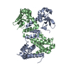 8u0gC  8u1jC C: citing same article ( |
|---|---|
| Experimental dataset #1 | Data reference: 10.5281/zenodo.10022358 / Data set type: diffraction image data / Metadata reference: 10.5281/zenodo.10022358 |
-Links
 PDBj
PDBj- Assembly
| Deposited unit | 
| ||||||||||||
|---|---|---|---|---|---|---|---|---|---|---|---|---|---|
| 1 |
| ||||||||||||
| Unit cell |
|
-Components
| #1: Protein | Mass: 25407.971 Da / Num. of mol.: 2 / Mutation: L131M, L184M Source method: isolated from a genetically manipulated source Source: (gene. exp.) Deinococcus radiodurans (radioresistant)Plasmid: pDEST-527 / Details (production host): TEV-cleavable 6xHis tag (N-term) / Production host: #2: Chemical | ChemComp-SO4 /   Mass: 96.063 Da / Num. of mol.: 4 / Source method: obtained synthetically / Formula: SO4 Mass: 96.063 Da / Num. of mol.: 4 / Source method: obtained synthetically / Formula: SO4#3: Water | ChemComp-HOH / |  Mass: 18.015 Da / Num. of mol.: 40 / Source method: isolated from a natural source / Formula: H2O Mass: 18.015 Da / Num. of mol.: 40 / Source method: isolated from a natural source / Formula: H2OHas ligand of interest | N | Has protein modification | Y | |
|---|
-Experimental details
-Experiment
| Experiment | Method: X-RAY DIFFRACTION / Number of used crystals: 1 |
|---|
- Sample preparation
| Crystal | Density Matthews: 2.84 Å3/Da / Density % sol: 56.63 % / Description: Elongated square bipyramid |
|---|---|
| Crystal grow | Temperature: 293 K / Method: vapor diffusion, hanging drop / pH: 7.5 Details: 250 uM DdrC 140 mM Sodium sulfate 14 mM Sodium citrate 3.6% (w/v) PEG 8000 4.3% (v/v) Pentaerythritol ethoxylate (3/4 EO/OH) 18 mM HEPES/NaOH, pH 7.5 |
-Data collection
| Diffraction | Mean temperature: 100 K / Ambient temp details: Nitrogen Cryo-stream / Serial crystal experiment: N |
|---|---|
| Diffraction source | Source: SYNCHROTRON / Site: CLSI / Beamline: 08B1-1 / Wavelength: 0.9795 Å |
| Detector | Type: DECTRIS PILATUS3 S 6M / Detector: PIXEL / Date: Jan 13, 2022 Details: Incorporated white beam slits (WBS), a vertical collimating mirror (VCM), a double-crystal monochromator (DCM) / double-multilayer monochromator (DMM), and toroidal focusing mirror |
| Radiation | Monochromator: KOHZU Si(111) double crystal monochromator / Protocol: SINGLE WAVELENGTH / Monochromatic (M) / Laue (L): M / Scattering type: x-ray |
| Radiation wavelength | Wavelength: 0.9795 Å / Relative weight: 1 |
| Reflection | Resolution: 2.239→66.698 Å / Num. obs: 26343 / % possible obs: 96.8 % / Redundancy: 10.1 % / Biso Wilson estimate: 54.41 Å2 / CC1/2: 0.998 / Rmerge(I) obs: 0.055 / Rpim(I) all: 0.023 / Rrim(I) all: 0.073 / Net I/σ(I): 19.1 |
| Reflection shell | Resolution: 2.239→2.278 Å / Redundancy: 5.2 % / Rmerge(I) obs: 0.706 / Mean I/σ(I) obs: 2.3 / Num. unique obs: 1335 / CC1/2: 0.631 / Rpim(I) all: 0.392 / Rrim(I) all: 0.899 / % possible all: 99.9 |
- Processing
| Software |
| |||||||||||||||||||||||||||||||||||||||||||||||||||||||||||||||||||||||||||||||||||||||||||||||||||||||||||||||||||||||||||||||||||||
|---|---|---|---|---|---|---|---|---|---|---|---|---|---|---|---|---|---|---|---|---|---|---|---|---|---|---|---|---|---|---|---|---|---|---|---|---|---|---|---|---|---|---|---|---|---|---|---|---|---|---|---|---|---|---|---|---|---|---|---|---|---|---|---|---|---|---|---|---|---|---|---|---|---|---|---|---|---|---|---|---|---|---|---|---|---|---|---|---|---|---|---|---|---|---|---|---|---|---|---|---|---|---|---|---|---|---|---|---|---|---|---|---|---|---|---|---|---|---|---|---|---|---|---|---|---|---|---|---|---|---|---|---|---|---|
| Refinement | Method to determine structure: SAD / Resolution: 2.24→59.3 Å / SU ML: 0.3187 / Cross valid method: FREE R-VALUE / σ(F): 1.34 / Phase error: 30.0333 Stereochemistry target values: GeoStd + Monomer Library + CDL v1.2
| |||||||||||||||||||||||||||||||||||||||||||||||||||||||||||||||||||||||||||||||||||||||||||||||||||||||||||||||||||||||||||||||||||||
| Solvent computation | Shrinkage radii: 0.9 Å / VDW probe radii: 1.11 Å / Solvent model: FLAT BULK SOLVENT MODEL | |||||||||||||||||||||||||||||||||||||||||||||||||||||||||||||||||||||||||||||||||||||||||||||||||||||||||||||||||||||||||||||||||||||
| Displacement parameters | Biso mean: 66.54 Å2 | |||||||||||||||||||||||||||||||||||||||||||||||||||||||||||||||||||||||||||||||||||||||||||||||||||||||||||||||||||||||||||||||||||||
| Refinement step | Cycle: LAST / Resolution: 2.24→59.3 Å
| |||||||||||||||||||||||||||||||||||||||||||||||||||||||||||||||||||||||||||||||||||||||||||||||||||||||||||||||||||||||||||||||||||||
| Refine LS restraints |
| |||||||||||||||||||||||||||||||||||||||||||||||||||||||||||||||||||||||||||||||||||||||||||||||||||||||||||||||||||||||||||||||||||||
| LS refinement shell |
|
