 Movie
Movie Controller


+ Open data

- Basic information
| Entry | Database: PDB / ID: 8tuk | |||||||||||||||
|---|---|---|---|---|---|---|---|---|---|---|---|---|---|---|---|---|
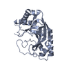
| Title | Alvinella ASCC1 KH and Phosphodiesterase/Ligase Domain | |||||||||||||||
 Components Components | Activating signal cointegrator 1 complex subunit 1 | |||||||||||||||
 Keywords Keywords | RNA BINDING PROTEIN / alkylation response / RNA damage / KH domain / phosphoesterase domain / RNA ligase domain | |||||||||||||||
| Function / homology | IMIDAZOLE Function and homology information Function and homology information | |||||||||||||||
| Biological species |  Alvinella pompejana (invertebrata) Alvinella pompejana (invertebrata) | |||||||||||||||
| Method |  X-RAY DIFFRACTION / SYNCHROTRON / MOLECULAR REPLACEMENT / Resolution: 1.15 Å X-RAY DIFFRACTION / SYNCHROTRON / MOLECULAR REPLACEMENT / Resolution: 1.15 Å | |||||||||||||||
 Authors Authors | Tsutakawa, S.E. / Tainer, J.A. / Arvai, A.S. / Chinnam, N.B. | |||||||||||||||
| Funding support |  United States, 4items United States, 4items
| |||||||||||||||
 Citation Citation | Journal: J.Biol.Chem. / Year: 2024 Title: ASCC1 structures and bioinformatics reveal a novel helix-clasp-helix RNA-binding motif linked to a two-histidine phosphodiesterase. Authors: Chinnam, N.B. / Thapar, R. / Arvai, A.S. / Sarker, A.H. / Soll, J.M. / Paul, T. / Syed, A. / Rosenberg, D.J. / Hammel, M. / Bacolla, A. / Katsonis, P. / Asthana, A. / Tsai, M.S. / Ivanov, I. ...Authors: Chinnam, N.B. / Thapar, R. / Arvai, A.S. / Sarker, A.H. / Soll, J.M. / Paul, T. / Syed, A. / Rosenberg, D.J. / Hammel, M. / Bacolla, A. / Katsonis, P. / Asthana, A. / Tsai, M.S. / Ivanov, I. / Lichtarge, O. / Silverman, R.H. / Mosammaparast, N. / Tsutakawa, S.E. / Tainer, J.A. #1: Journal: Proteins / Year: 2021Title: Target highlights in CASP14: Analysis of models by structure providers. Authors: Alexander, L.T. / Lepore, R. / Kryshtafovych, A. / Adamopoulos, A. / Alahuhta, M. / Arvin, A.M. / Bomble, Y.J. / Bottcher, B. / Breyton, C. / Chiarini, V. / Chinnam, N.B. / Chiu, W. / ...Authors: Alexander, L.T. / Lepore, R. / Kryshtafovych, A. / Adamopoulos, A. / Alahuhta, M. / Arvin, A.M. / Bomble, Y.J. / Bottcher, B. / Breyton, C. / Chiarini, V. / Chinnam, N.B. / Chiu, W. / Fidelis, K. / Grinter, R. / Gupta, G.D. / Hartmann, M.D. / Hayes, C.S. / Heidebrecht, T. / Ilari, A. / Joachimiak, A. / Kim, Y. / Linares, R. / Lovering, A.L. / Lunin, V.V. / Lupas, A.N. / Makbul, C. / Michalska, K. / Moult, J. / Mukherjee, P.K. / Nutt, W.S. / Oliver, S.L. / Perrakis, A. / Stols, L. / Tainer, J.A. / Topf, M. / Tsutakawa, S.E. / Valdivia-Delgado, M. / Schwede, T. #2: Journal: Methods Mol.Biol. / Year: 2022 Title: Universally Accessible Structural Data on Macromolecular Conformation, Assembly, and Dynamics by Small Angle X-Ray Scattering for DNA Repair Insights. Authors: Chinnam, N.B. / Syed, A. / Burnett, K.H. / Hura, G.L. / Tainer, J.A. / Tsutakawa, S.E. #3: Journal: Methods Enzymol. / Year: 2023 Title: Combining small angle X-ray scattering (SAXS) with protein structure predictions to characterize conformations in solution. Authors: Chinnam, N.B. / Syed, A. / Hura, G.L. / Hammel, M. / Tainer, J.A. / Tsutakawa, S.E. | |||||||||||||||
| History |
|
- Structure visualization
| Structure viewer | Molecule: MolmilJmol/JSmol |
|---|
- Downloads & links
-Download
| PDBx/mmCIF format | 8tuk.cif.gz | 216.6 KB | Display | PDBx/mmCIF format |
|---|---|---|---|---|
| PDB format | pdb8tuk.ent.gz | 146.7 KB | Display | PDB format |
| PDBx/mmJSON format | 8tuk.json.gz | Tree view | PDBx/mmJSON format | |
| Others |  Other downloads Other downloads |
-Validation report
| Arichive directory | https://data.pdbj.org/pub/pdb/validation_reports/tu/8tukftp://data.pdbj.org/pub/pdb/validation_reports/tu/8tuk | HTTPS FTP |
|---|
-Related structure data
-Links
 PDBj
PDBj


- Assembly
| Deposited unit | 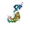
| ||||||||||||
|---|---|---|---|---|---|---|---|---|---|---|---|---|---|
| 1 |
| ||||||||||||
| Unit cell |
|
-Components
| #1: Protein | Mass: 36619.336 Da / Num. of mol.: 1 Source method: isolated from a genetically manipulated source Source: (gene. exp.) Alvinella pompejana (invertebrata) / Gene: 2696536 / Production host:  | ||||||
|---|---|---|---|---|---|---|---|
| #2: Chemical |   Mass: 69.085 Da / Num. of mol.: 2 / Source method: obtained synthetically / Formula: C3H5N2 Mass: 69.085 Da / Num. of mol.: 2 / Source method: obtained synthetically / Formula: C3H5N2#3: Chemical | ChemComp-EDO /   Mass: 62.068 Da / Num. of mol.: 5 / Source method: obtained synthetically / Formula: C2H6O2 Mass: 62.068 Da / Num. of mol.: 5 / Source method: obtained synthetically / Formula: C2H6O2#4: Water | ChemComp-HOH / |  Mass: 18.015 Da / Num. of mol.: 431 / Source method: isolated from a natural source / Formula: H2O Mass: 18.015 Da / Num. of mol.: 431 / Source method: isolated from a natural source / Formula: H2OHas ligand of interest | N | |
-Experimental details
-Experiment
| Experiment | Method: X-RAY DIFFRACTION / Number of used crystals: 1 |
|---|
- Sample preparation
| Crystal | Density Matthews: 2.07 Å3/Da / Density % sol: 40.59 % |
|---|---|
| Crystal grow | Temperature: 277 K / Method: vapor diffusion, hanging drop / pH: 5 Details: 15% MPEG 2K, 200 mM, 200 mM I/M pH 5.0. 2.5% KCl (saturated), 0.6% BME. For cryo protection, crystals for about 2 seconds in 15% MPEG 2K, 200 mM I/M pH 5.0, 2.5% KCl (saturated), 60% ...Details: 15% MPEG 2K, 200 mM, 200 mM I/M pH 5.0. 2.5% KCl (saturated), 0.6% BME. For cryo protection, crystals for about 2 seconds in 15% MPEG 2K, 200 mM I/M pH 5.0, 2.5% KCl (saturated), 60% ethylene glycol was mixed 1:2 (cryo:reservoir) Temp details: 15 deg celcius |
-Data collection
| Diffraction | Mean temperature: 100 K / Serial crystal experiment: N |
|---|---|
| Diffraction source | Source: SYNCHROTRON / Site: SSRL / Beamline: BL9-2 / Wavelength: 0.97946 Å |
| Detector | Type: DECTRIS PILATUS 6M / Detector: PIXEL / Date: Feb 27, 2020 |
| Radiation | Protocol: SINGLE WAVELENGTH / Monochromatic (M) / Laue (L): M / Scattering type: x-ray |
| Radiation wavelength | Wavelength: 0.97946 Å / Relative weight: 1 |
| Reflection | Resolution: 1.15→3.44 Å / Num. obs: 101451 / % possible obs: 95.7 % / Redundancy: 6.5 % / Biso Wilson estimate: 15.76 Å2 / CC1/2: 0.999 / Net I/σ(I): 6.5 |
| Reflection shell | Resolution: 1.15→1.22 Å / Redundancy: 5 % / Mean I/σ(I) obs: 5 / Num. unique obs: 13759 / CC1/2: 0.83 / % possible all: 80.8 |
- Processing
| Software |
| |||||||||||||||||||||||||||||||||||||||||||||||||||||||||||||||||||||||||||||||||||||||||||||||||||||||||
|---|---|---|---|---|---|---|---|---|---|---|---|---|---|---|---|---|---|---|---|---|---|---|---|---|---|---|---|---|---|---|---|---|---|---|---|---|---|---|---|---|---|---|---|---|---|---|---|---|---|---|---|---|---|---|---|---|---|---|---|---|---|---|---|---|---|---|---|---|---|---|---|---|---|---|---|---|---|---|---|---|---|---|---|---|---|---|---|---|---|---|---|---|---|---|---|---|---|---|---|---|---|---|---|---|---|---|
| Refinement | Method to determine structure: MOLECULAR REPLACEMENT / Resolution: 1.15→3.44 Å / SU ML: 0.0928 / Cross valid method: FREE R-VALUE / σ(F): 0.09 / Phase error: 18.7934 Stereochemistry target values: GeoStd + Monomer Library + CDL v1.2
| |||||||||||||||||||||||||||||||||||||||||||||||||||||||||||||||||||||||||||||||||||||||||||||||||||||||||
| Solvent computation | Shrinkage radii: 0.9 Å / VDW probe radii: 1.11 Å / Solvent model: FLAT BULK SOLVENT MODEL | |||||||||||||||||||||||||||||||||||||||||||||||||||||||||||||||||||||||||||||||||||||||||||||||||||||||||
| Displacement parameters | Biso mean: 29.48 Å2 | |||||||||||||||||||||||||||||||||||||||||||||||||||||||||||||||||||||||||||||||||||||||||||||||||||||||||
| Refinement step | Cycle: LAST / Resolution: 1.15→3.44 Å
| |||||||||||||||||||||||||||||||||||||||||||||||||||||||||||||||||||||||||||||||||||||||||||||||||||||||||
| Refine LS restraints |
| |||||||||||||||||||||||||||||||||||||||||||||||||||||||||||||||||||||||||||||||||||||||||||||||||||||||||
| LS refinement shell |
|
